42 eye cross section diagram
Human eye anatomy cross section medical diagram, vector flat isolated illustration. Structure of healthy eye and with glaucoma, cataract, hyperopia, myopia diseases and disorders. Human eye anatomy, right eye viewed from above Vector Illustration Of Human eye anatomy, right eye viewed from above choroid stock illustrations. Human eye cross section anatomy diagram - download this royalty free Vector in seconds. No membership needed. Human eye cross section anatomy diagram with all parts anatomical structure for medical science education and health care. | CanStock
Müllers muscle: Sympathetically-innervated muscle extending from levator palpebrae superioris to top of tarsal plate. Elevates eyelid, but not nearly as much as levator does. Impaired in Horner Syndrome, but ptosis is mild because Müller's muscle is a weak elevator. Orbital septum: Collagenous curtain connecting frontal bone and upper lid tarsus.

Eye cross section diagram
Figure 5. Waveform and resultant eye diagram with 75% eye crossing percentage Bit Period The bit period is a measure of the horizontal opening of an eye diagram at the crossing points of the eye and is usually measured in picoseconds for a high speed digital signal (i.e., 200 ps is used for a 5 Gbps signal). The data rate is the the outer eye as we see it with all parts labeled lateral view of the eyeball in the skull top view of the eyeball in the skull diagram of the visual field large central illustration of a lateral cross section of the eye medial cross section of the eye close up views of the: anterior chamber angle lens retina fundus macula lutea Made in the USA. Eye Cross Section Labeled Diagram. An eye cut saggital labeled diagram. Human eye anatomy. Cartoon simple illustration for medical atlas or educational textbook. Cross-section of an eyes. Human eye anatomy. Abstract polygonal light of human eye anatomy. Business wireframe mesh spheres from flying debris. Eye cross section concept.
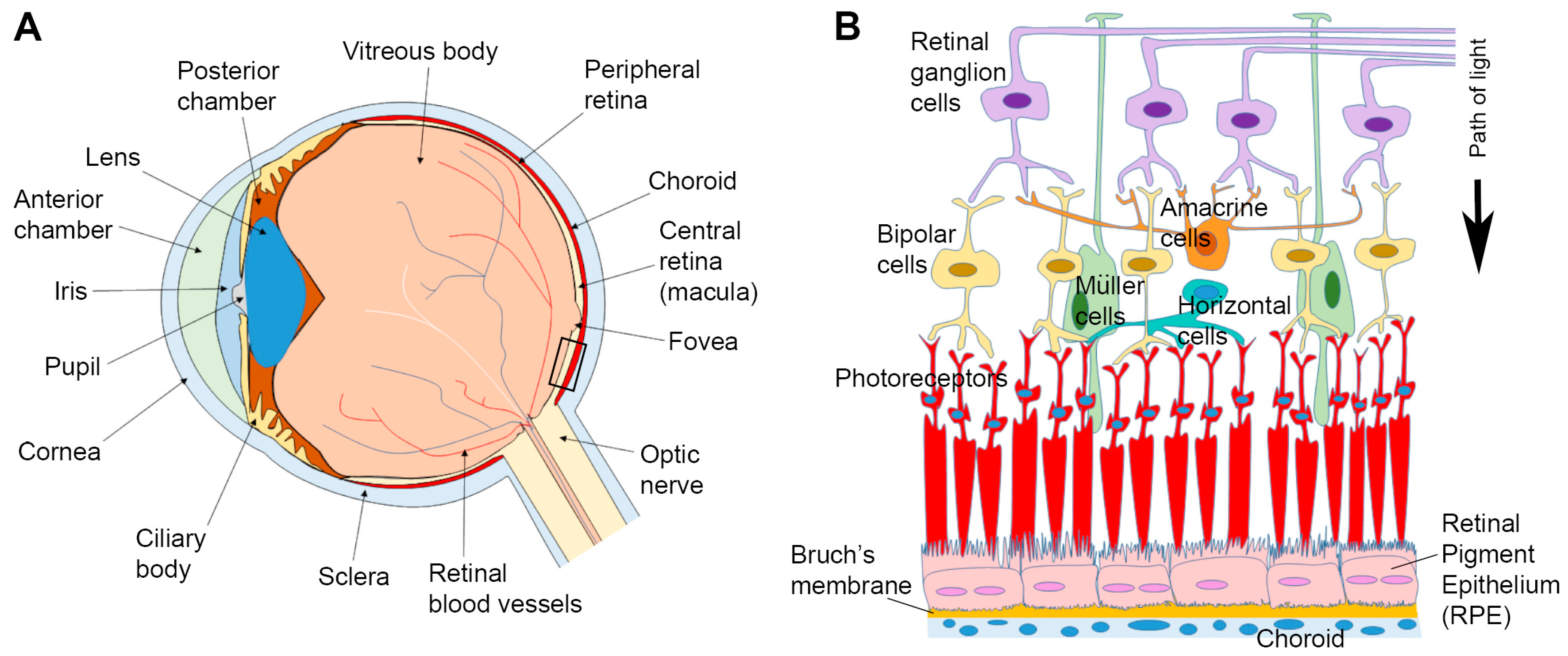
Eye cross section diagram. The arrangement of retinal cells is shown in a cross section. Vector diagram for your design, educational, biological, science and medical use human eye anatomy stock illustrations. Eye Cross section Anatomy (with name) Eye Cross section Anatomy (with name) human eye anatomy stock illustrations ... Diagrams are available in two file formats: JPG, AI (Adobe Illustrator). ... You're viewing: Human Eye in Cross Section $ 0.00. Select options Answer The normal is an imaginary line drawn on a ray diagram perpendicular to, so at a right angle (90 degrees), to the boundary between two media. ... Eye in Cross Section. Click on a label to display the definition. Tap on the image or pinch out and pinch in to resize the image. Conjunctiva: mucous membrane covering anterior eye. bulbar conjunctiva covers sclera. palpebral conjunctiva covers inner surface of eyelids. keeps cornea moist and fends off infection. Cross-section anatomical diagram of a cataract developing in an eye. Soft Foam Cross-Section Eye Model. Check for more insights of every cell unit of a leaf. Cross section through the maxillary sinus. Choose an option 400 pixels 800 pixels Clear. Evoke the excitement of exploration with this Edraw leaf cross section science diagram template.
Above is a human eye cross section diagram I sketched out for a paper I was working on at the time. Tagged art, cross section, diagram, eye, human eye, science, sketch. Oct 15 2017. Leave a comment. Drawing, Ink, Paper, Pencil. Opossum. An opossum illustration I made for a friend. Download a diagram and explanation of the cross section of the human eye. Download this Free Vector about Diagram showing cross section of human eye, and discover more than 20 Million Professional Graphic Resources on Freepik. #freepik #vector #background #people #education Download scientific diagram | Structure of a generalized reptilian eye. A, Cross section of a lizard eye; B, Corneal section of an eye. Abbreviations: Bm, Brücke's muscle; c, cornea; Cm ...
Cross section through the tongue and C2: Diagram This cross-section has the exact same orientation as the previous one. The posterior landmark is provided by the second cervical vertebra (axis) while the anterior one is provided by the tongue. However, there are quite a few differences between them. 6,636 human eye diagram stock photos, vectors, and illustrations are available royalty-free. See human eye diagram stock video clips. of 67. diagram of the eye diagram of eyeball cross section of human eye section of human eye cross section eye cross section of eye anatomy of the eye eye parts human eye diagram of eye. The diagrams below show cross sections of the human eyeball. As we journey through the different structures, refer to the diagrams to quickly digest the content on this page. Our eyeballs are fairly round organs cushioned by fatty tissues and they sit in two bony sockets inside the skull. This helps to protect our eyes from injury. Human eye anatomy with cross section of eye diagram illustration; Ophthalmology Infographic. Human Eye Anatomy Structure Banner with Cross Section of Vision Organ. UVB, UVA Rays Protection. Wet and Dry Macular Degeneration. Realistic Vector Illustration, Copy Space.
Dysfunctional Lens Syndrome Treatment in East Hanover, NJ
Browse 405 eye cross section stock illustrations and vector graphics available royalty-free, or search for glaucoma or human eye to find more great stock images and vector art. human eye diagram - eye cross section stock illustrations. eye anatomy, artwork - eye cross section stock illustrations.

Lateral Cross-Sectional View of the Eye - TrialExhibits Inc.
Diagram showing cross section of human eye. Holding a silicone anatomy of an eye in Hand. Diagram showing parts of human eye illustration. Corneal abrasion in the human eye. eye vein system x ray angiography vector design ...
Eye Anatomy Kogarah | Cross Section of Eye Wollongong | Sydney
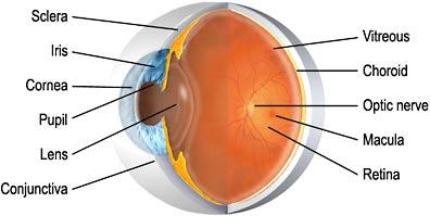
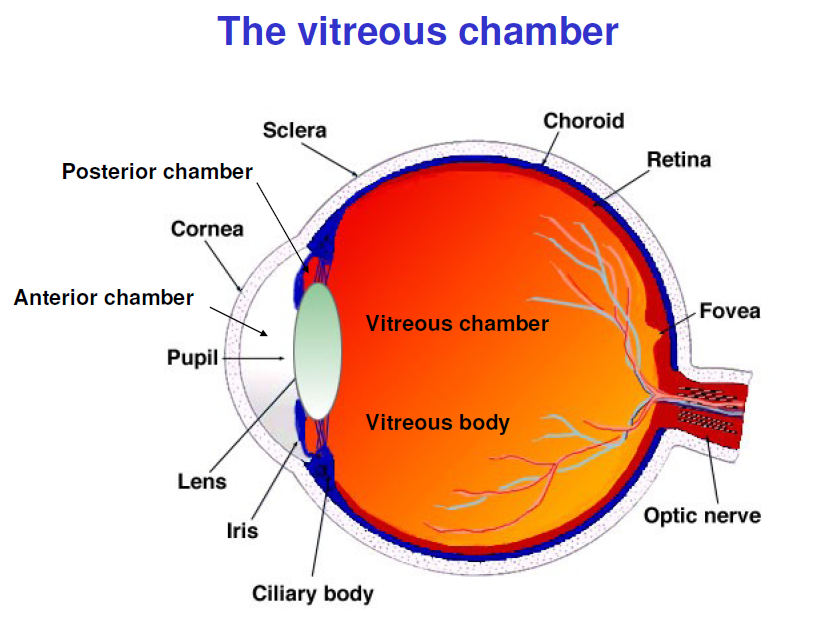
Nov 19, 2021 · A, Cross section diagram of the eye with emphasis on the anatomical features of the vitreous. The vitreous is most firmly attached to the retina at the vitreous base, and it also has adhesions at the optic nerve, along vessels, at the fovea, and to the posterior lens capsule. A prominent area of liquefaction of the premacular vitreous gel is called the premacular bursa, or precortical vitreous pocket.

The S.Y.J.R, a 7-year perspective, Part One, at Firbeck, Maltby & Brookhouse - various 0273->9269
Download this Free Vector about Diagram showing cross section of human eye, and discover more than 20 Million Professional Graphic Resources on Freepik. #freepik #Vector #Education #Character #Chart

A cross-section diagram of the eye | Nervous system ...
This Stock Illustration, whose title is "Structure cross section of eye"[19950790], includes tags of eyes, eye, cross-section diagram. The author of this item is koti (No.295268). Sizes from S to XL are available and the price starts from US$5.00.

Posterior eye anatomy. (A) Cross-section schematic of a ...
Eye anatomy blank worksheet, printable test illustrations. Brain and eye anatomical cross section diagrams. School lesson activity educational workbook page.

Age Related Macular Degeneration - Nottingham | West ...
Lobster eyes—brilliant geometric design Lobster eyes, X-ray telescopes, and microchips. by Jonathan Sarfati. Diagram showing how the lobster eye focuses light. Adapted from Denton. 5 Click for larger view. The eye of a lobster (and some other 10-legged crustaceans 1 including shrimps and prawns) shows a remarkable geometry not found elsewhere in nature—it has tiny facets that are perfectly ...

Eye
cross section of human eye. - retina diagram stock illustrations old engraved illustration of eye infections and diseases - retina diagram stock pictures, royalty-free photos & images old engraved illustration of eye infections and diseases, cataracta capsularis (turbidity of the lens capsule after a cataract surgery) - retina diagram stock ...
Vertical Section of the Eyeball | ClipArt ETC
Use this human eye diagram to help you to teach children about the eye. This cross-section has been illustrated using clear black and white lines - it's ideal to use in colouring and labelling activities. The human eye has many parts that work together to produce vision: such as the pupil, iris and sclera.
Cells | Free Full-Text | Impaired Cargo Clearance in the ...
Structure of eye cross section - Stock Illustration(No.19951005). Find images exactly you are looking for from more than 67,400,000 of royalty-free stock photos, illustrations, and vectors. Download and enjoy fresh & incredible images added every day.

Schematic cross-section of the human eye. Source Wikipedia ...
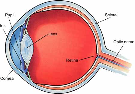
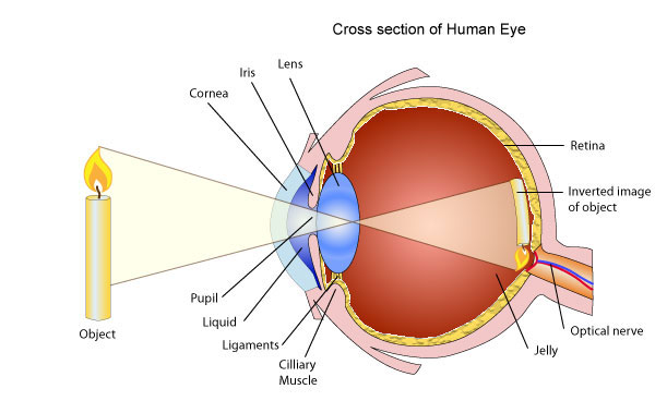
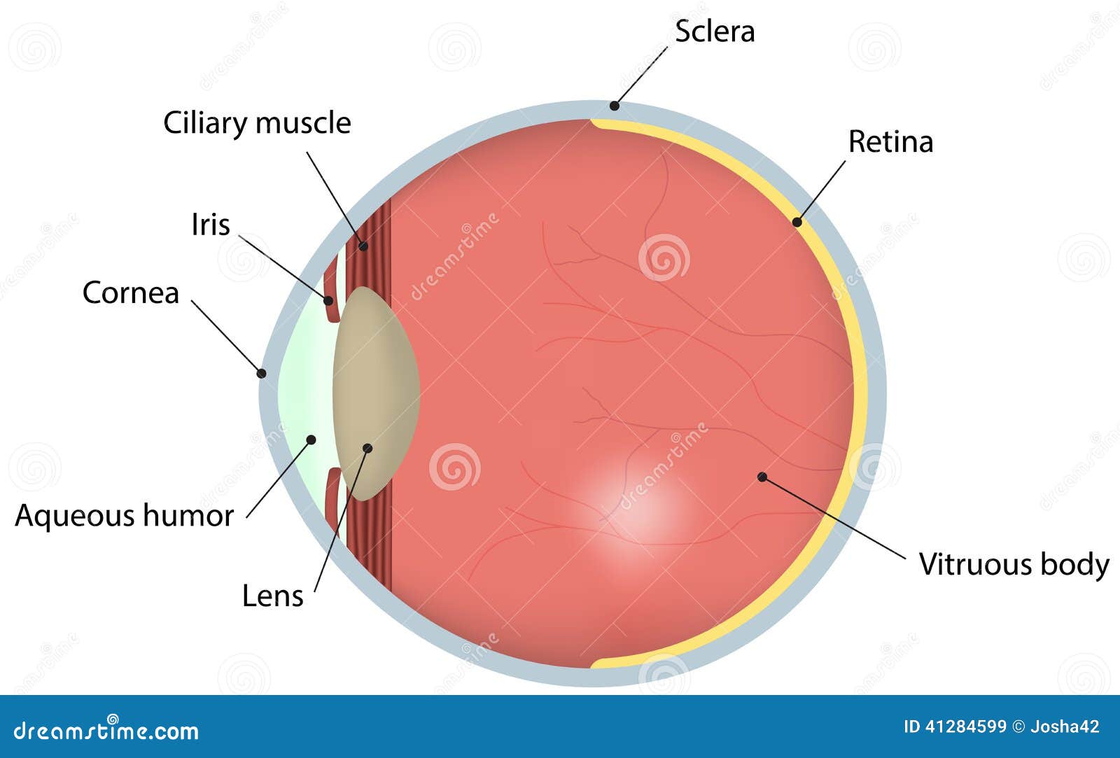
Jan 28, 2015 · Eye Cross-section. When light strikes the eye, the first part it reaches is the cornea, a dome positioned over the center of the eye. The cornea is clear and refracts, or bends, the light passing ...
Use of lenses for correcting vision - Pass My Exams: Easy ...
Eye Cross Section Labeled Diagram. An eye cut saggital labeled diagram. Human eye anatomy. Cartoon simple illustration for medical atlas or educational textbook. Cross-section of an eyes. Human eye anatomy. Abstract polygonal light of human eye anatomy. Business wireframe mesh spheres from flying debris. Eye cross section concept.

Human Eye Cross Section Eyeball 3D Model OBJ 3DS FBX C4D ...
the outer eye as we see it with all parts labeled lateral view of the eyeball in the skull top view of the eyeball in the skull diagram of the visual field large central illustration of a lateral cross section of the eye medial cross section of the eye close up views of the: anterior chamber angle lens retina fundus macula lutea Made in the USA.
1: Basic anatomy of the human eye, as seen through a cross ...
Figure 5. Waveform and resultant eye diagram with 75% eye crossing percentage Bit Period The bit period is a measure of the horizontal opening of an eye diagram at the crossing points of the eye and is usually measured in picoseconds for a high speed digital signal (i.e., 200 ps is used for a 5 Gbps signal). The data rate is the

Cross-sectional anatomy of the upper and lower eyelids ...

Cross section of a human eye anatomy. | Download ...
Human Eye Cross Section Normal Vision Stock Illustration ...

1: Human eye cross-sectional view with an example of a ...

Medical Stock Art, Anatomy of the Eye
The Brain and the Eye - How They Work Together - Discovery ...

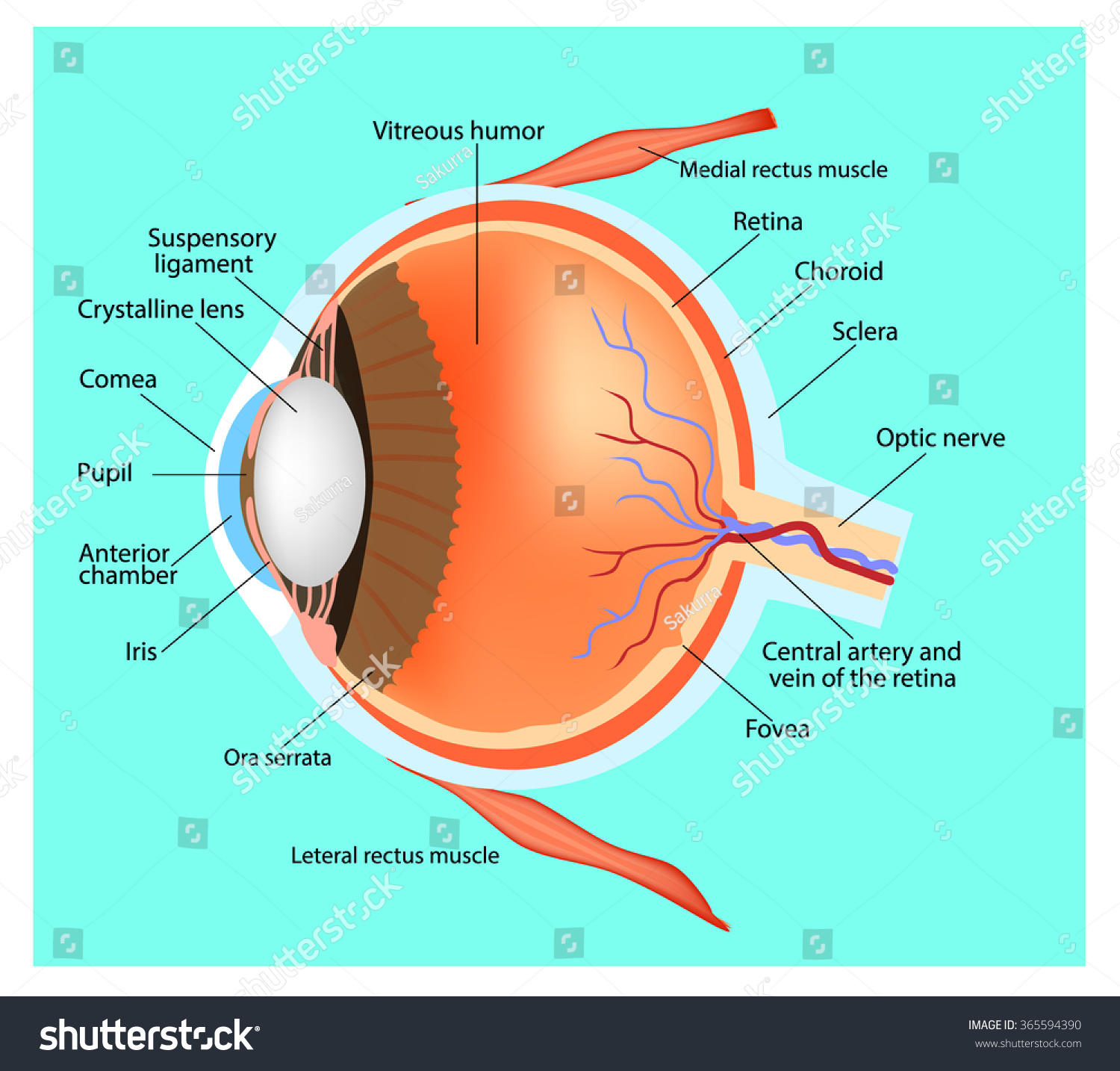
The main components of a human eye in horizontal cross ...

(a) Cross-section of the human eye. (b) Expanded view of ...

The cross-section of the human eye. | Download Scientific ...

A cross-section anatomy of the human eye. | Eye anatomy ...

A schematic cross section of the eye. The eye is made up ...

diagram of eye
Gross Anatomy of the Eye by Helga Kolb - Webvision

The London Eye

Cross-section through the human eye. Diagram by the author ...
Eye Cross Section Labeled Diagram Stock Vector ...
Neuro Lecture I Flashcards | Easy Notecards

O1 Eye Cross Section Diagram.jpg

Cross-section of a human eye with an expanded view of the ...

Hiking in autumn

Diagram Of The Eye Blind Spot - Diagramaica

crispy london

1 Simplified diagram of a cross section of the human eye ...

Cross-section of the human eye. | Download Scientific Diagram
Biofluid Mechanics

Chorioretinal anatomy. A: Cross-section of human eye. B ...
Structure Eye Human Eye Cross Section Stock Vector ...

Chicken Mummy Recipe
0 Response to "42 eye cross section diagram"
Post a Comment