41 meiotic division beads diagram
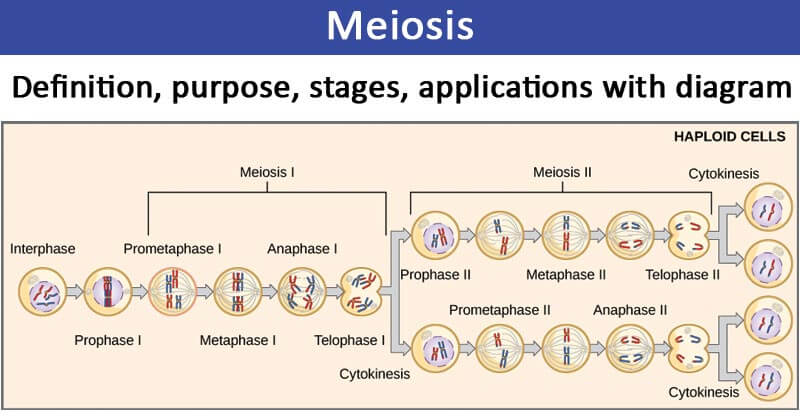
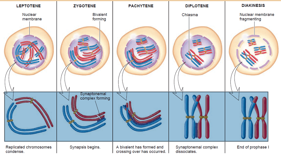
Junqueira's Basic Histology Text and Atlas, 14th Edition Meiotic Division of Cell (With Diagram) In this article we will discuss about the meiotic division of a cell. The meiotic division includes two complete divisions of a diploid cell resulting into four haploid nuclei. The first meiotic division includes a long prophase in which the homologous chromosomes become closely associated to each other ...
Explore this photo album by Darrietta Lee on Flickr!
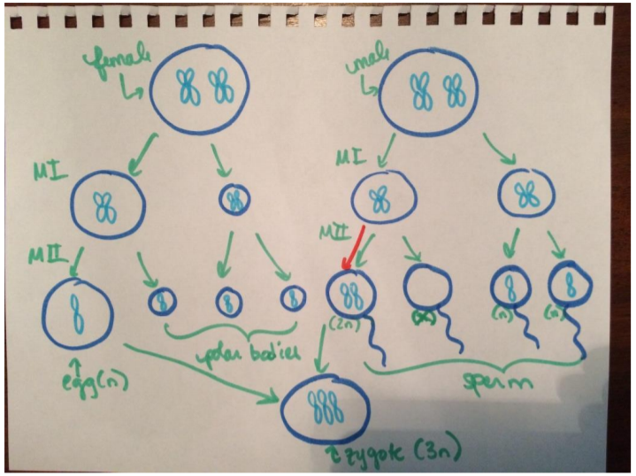
Meiotic division beads diagram
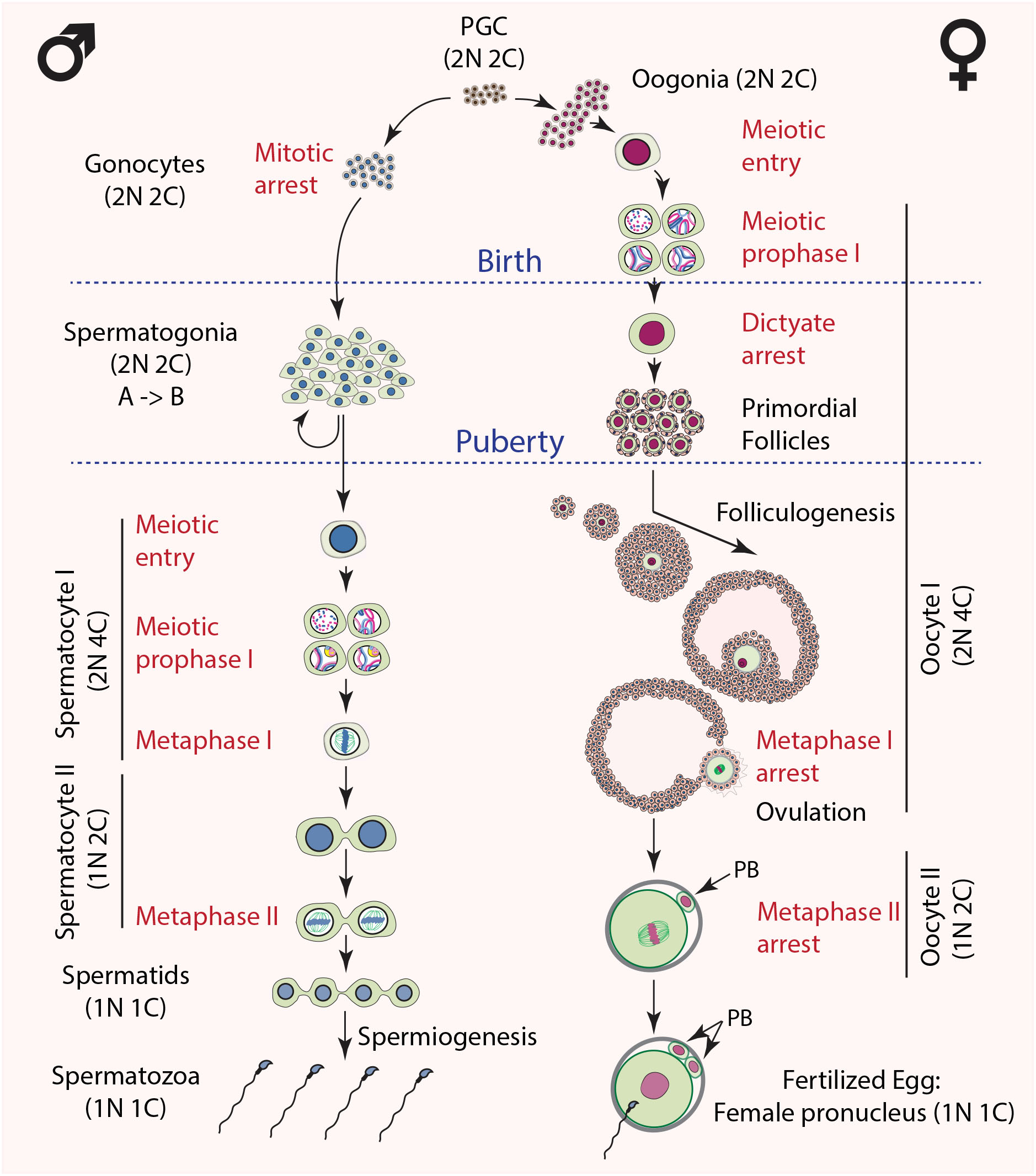
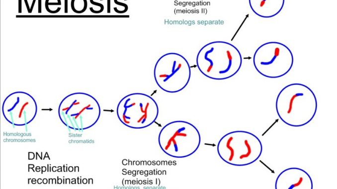
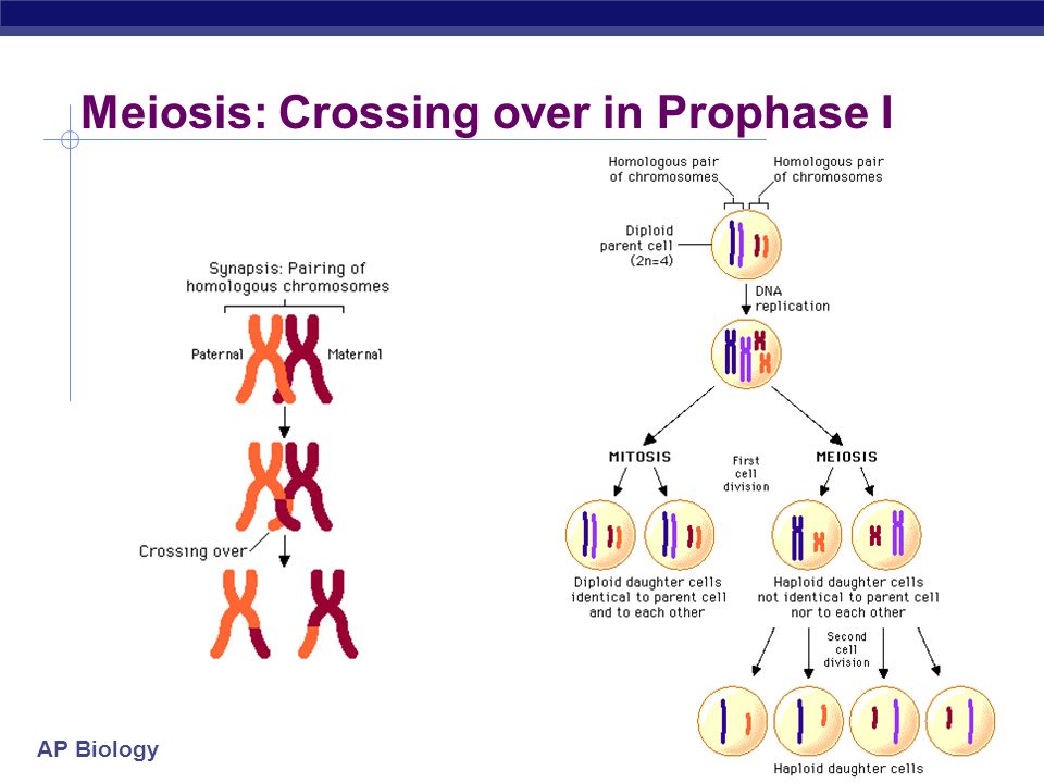
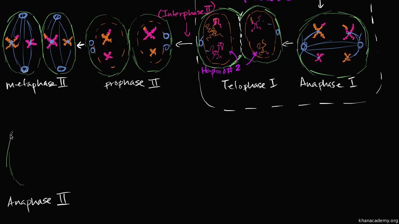
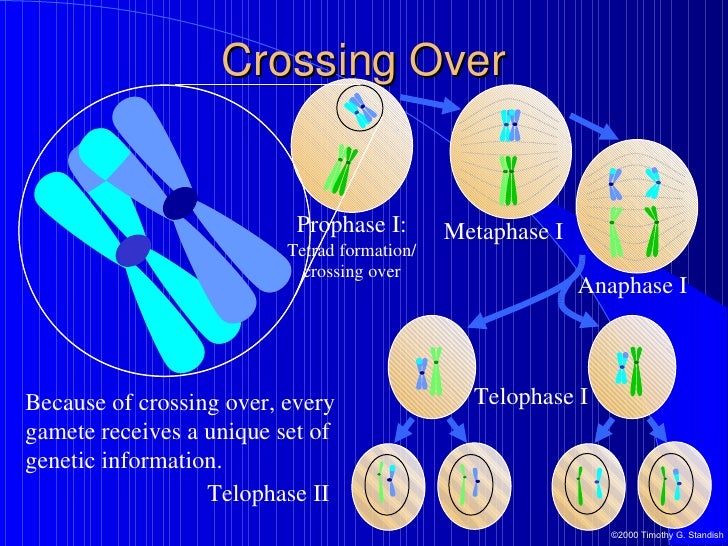
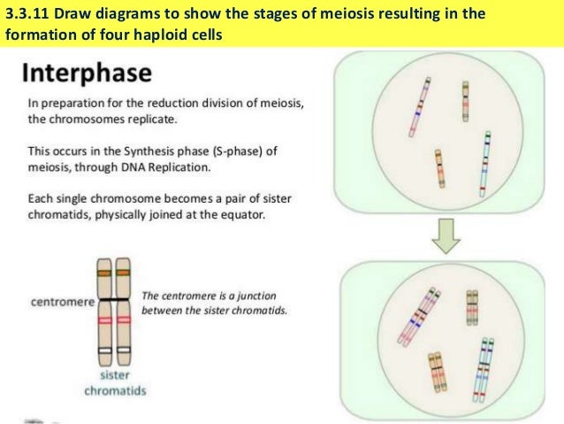
Meiotic cell division in males always results in four cells that become sperm. 2. Oogenesis. a. In the ovaries of human females, primary oocytes with 46 chromosomes undergo meiosis I to form two cells, each with 23 duplicated chromosomes. b. One of the cells, a secondary oocyte, receives almost all the cytoplasm; the other cell, a polar body, disintegrates or divides again. c. The secondary ... In preparation for the first division of meiosis, homologous chromosomes replicate and synapse. Like genes on the homologs align with each other. At this stage, segments of homologous chromosomes exchange linear segments of genetic material. This process is called recombination, or crossover, and it is a common genetic process. Because the genes are aligned during recombination, the gene order ... Insert picture here. Part 1 - Meiotic Division Beads Diagram. Prophase I. Metaphase I. Anaphase I.
Meiotic division beads diagram. pop beads. In lab, pop beads with magnetic centromeres are used to simulate chromosomes as they move through meiosis. The meiotic board used includes six circles: one on top; two in the middle and four at the bottom. These circles represent the chromosomal make up of the seven nuclei involved in one meiotic event. Trial 1 - Meiotic Division Beads Diagram. Part 2: Modeling Meiosis with Crossing Over Part 2 - Meiotic Division Beads Diagram: Image of page 3. Info icon This preview has intentionally blurred sections.Lab 4: Meiosis and Vertebrate Reproduction LAB SYNOPSIS: • Meiosis will be modeled using pop-beads. • The genetic diversity of gametes will ... 3:41Sara uses beads and diagrams to explain meiosis. View the attributions for all videos in the BIO106 playlist ...23 Feb 2015 · Uploaded by Elon TLT As we have studied, mitosis is the division of the nucleus of somatic (normal body) cells with the intent of making two exact copies of the parent cell. Meiosis ...3 pages
Biology questions and answers. Experiment 3: Following Chromosomal DNA Movement Through Meiosis Cell Cycle Division: Part 1 – Meiotic Bead Diagrams (Without Crossing Over) Prophase I Metaphase I Anaphase I Telophase I Prophase II Metaphase II Anaphase II Telophase II Cell Cycle Division: Part. 7:02A demo using lab beads to describe meiosis.15 Jul 2013 · Uploaded by Marsha Hay 5:27Kudos to my videographer, Amanda! This video is for an introductory biology class and is a simplified version of ...15 Mar 2016 · Uploaded by E Harrison Trial 2 Meiotic Division Beads Diagram Prophase I 4 Chromosomes Metaphase I 4 from BIOL MISC at Indiana University, Purdue University Indianapolis.
In biology, a good example of a liquid crystal is a meiotic spindle. A meiotic spindle has liquid-like properties, as it can fuse and deform and its molecular components turn over rapidly. The tubulin subunits in a spindle polymerize into microtubules, which order themselves by aligning along a common axis, and therefore also exhibit order Gatlin et al. 2010, Inoue 2008, Itabashi et al. 2009 ... a aa aaa aaaa aaacn aaah aaai aaas aab aabb aac aacc aace aachen aacom aacs aacsb aad aadvantage aae aaf aafp aag aah aai aaj aal aalborg aalib aaliyah aall aalto aam ... Cell Cycle Division: Part 2 – Meiotic Bead Diagrams (With Crossing Over) Prophase I: One chromosome from mother one from father come. together and wrap around each other so closely that portions of one switch. with portions of the other. They are also lined up along the middle. Anaphase II: The chromosomes are split in half and move to the. side Diagram the corresponding images for each stage in the sections titled “Trial 1 - Meiotic Division Beads Diagram”. Be sure to indicate the number of chromosomes present in each cell for each phase. 7. Disassemble the beads used in Part 1. You will need to recycle these beads for a second meiosis trial in Steps 8 - 13.
11:53Mr. Andersen uses chromosome beads to simulate both mitosis and meiosis. A brief discussion of gamete ...23 Nov 2010 · Uploaded by Bozeman Science
In cell biology, the spindle apparatus (or mitotic spindle) refers to the cytoskeletal structure of eukaryotic cells that forms during cell division to separate sister chromatids between daughter cells.It is referred to as the mitotic spindle during mitosis, a process that produces genetically identical daughter cells, or the meiotic spindle during meiosis, a process that produces gametes with ...
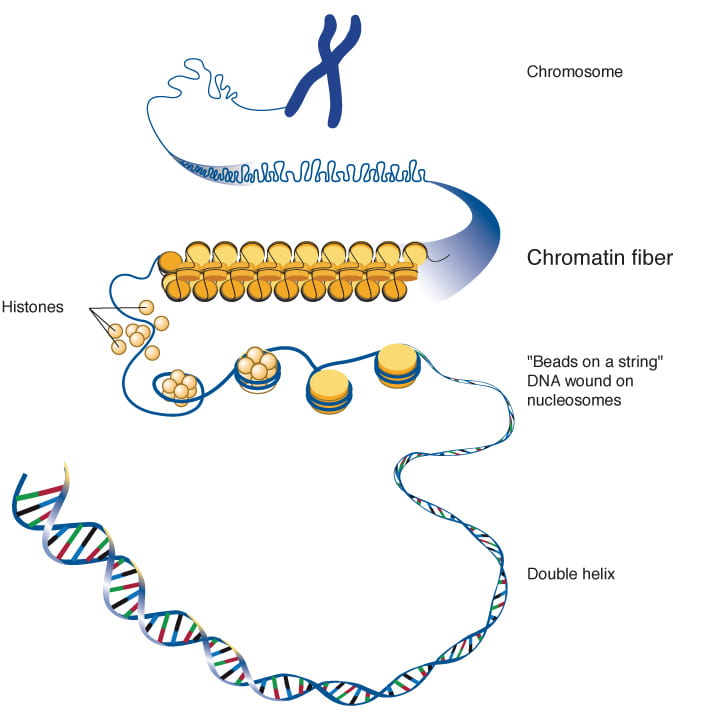
A chromosome is a long DNA molecule with part or all of the genetic material of an organism. Most eukaryotic chromosomes include packaging proteins called histones which, aided by chaperone proteins, bind to and condense the DNA molecule to maintain its integrity. These chromosomes display a complex three-dimensional structure, which plays a significant role in transcriptional regulation.
Diagram the corresponding images for each stage in the sections titled “Trial 1 - Meiotic Division Beads Diagram”. Be sure to indicate the number of chromosomes present in each cell for each phase. Disassemble the beads used in Part 1. You will need to recycle these beads for a second meiosis trial in Steps 7 - 12.
Biology I Lab Activity – Simulating Mitosis with “Pop Beads” Introduction: Mitosis is the process of one cell dividing to produce two new (daughter) cells (take a look at the diagram . Diagram the corresponding images for each stage in the section titled “Trial 2 - Meiotic Division Beads Diagram”.
7:42Next: · Lab 10: Part 1 - Meiosis bead demonstration · Mitosis Mastery using pipecleaner model · Phases of ...14 Jan 2013 · Uploaded by Kristen Hammer
Pipe cleaners, beads, or other appropriate materials for making After mitosis, the cytoplasm completes its division, forming two new cells. Label each phase and explain what is happening in each. Place both pipe cleaner halves though the bead. D: Cell Division - Pipe Cleaner Activity. Dump contents of bag onto table. Time. Activity #6. Hope-fully, they emerge from the activity with insight ...
100% (2 ratings) Meiosis comprises of two phases. In phase one the div …. View the full answer. Transcribed image text: Trial 2 - Meiotic Division Beads Diagram. Previous question Next question.
Insert picture here. Part 1 - Meiotic Division Beads Diagram. Prophase I. Metaphase I. Anaphase I.
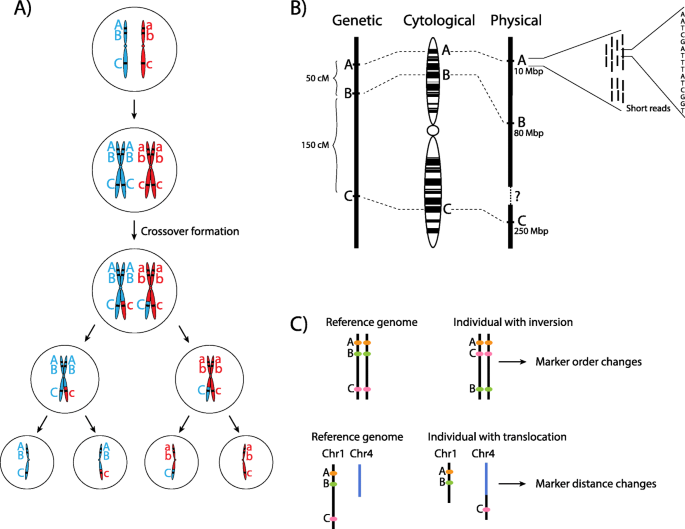
In preparation for the first division of meiosis, homologous chromosomes replicate and synapse. Like genes on the homologs align with each other. At this stage, segments of homologous chromosomes exchange linear segments of genetic material. This process is called recombination, or crossover, and it is a common genetic process. Because the genes are aligned during recombination, the gene order ...
Meiotic cell division in males always results in four cells that become sperm. 2. Oogenesis. a. In the ovaries of human females, primary oocytes with 46 chromosomes undergo meiosis I to form two cells, each with 23 duplicated chromosomes. b. One of the cells, a secondary oocyte, receives almost all the cytoplasm; the other cell, a polar body, disintegrates or divides again. c. The secondary ...

The Proteomic Landscape Of Centromeric Chromatin Reveals An Essential Role For The Ctf19ccan Complex In Meiotic Kinetochore Assembly Sciencedirect

Topoisomerases Modulate The Timing Of Meiotic Dna Breakage And Chromosome Morphogenesis In Saccharomyces Cerevisiae Genetics
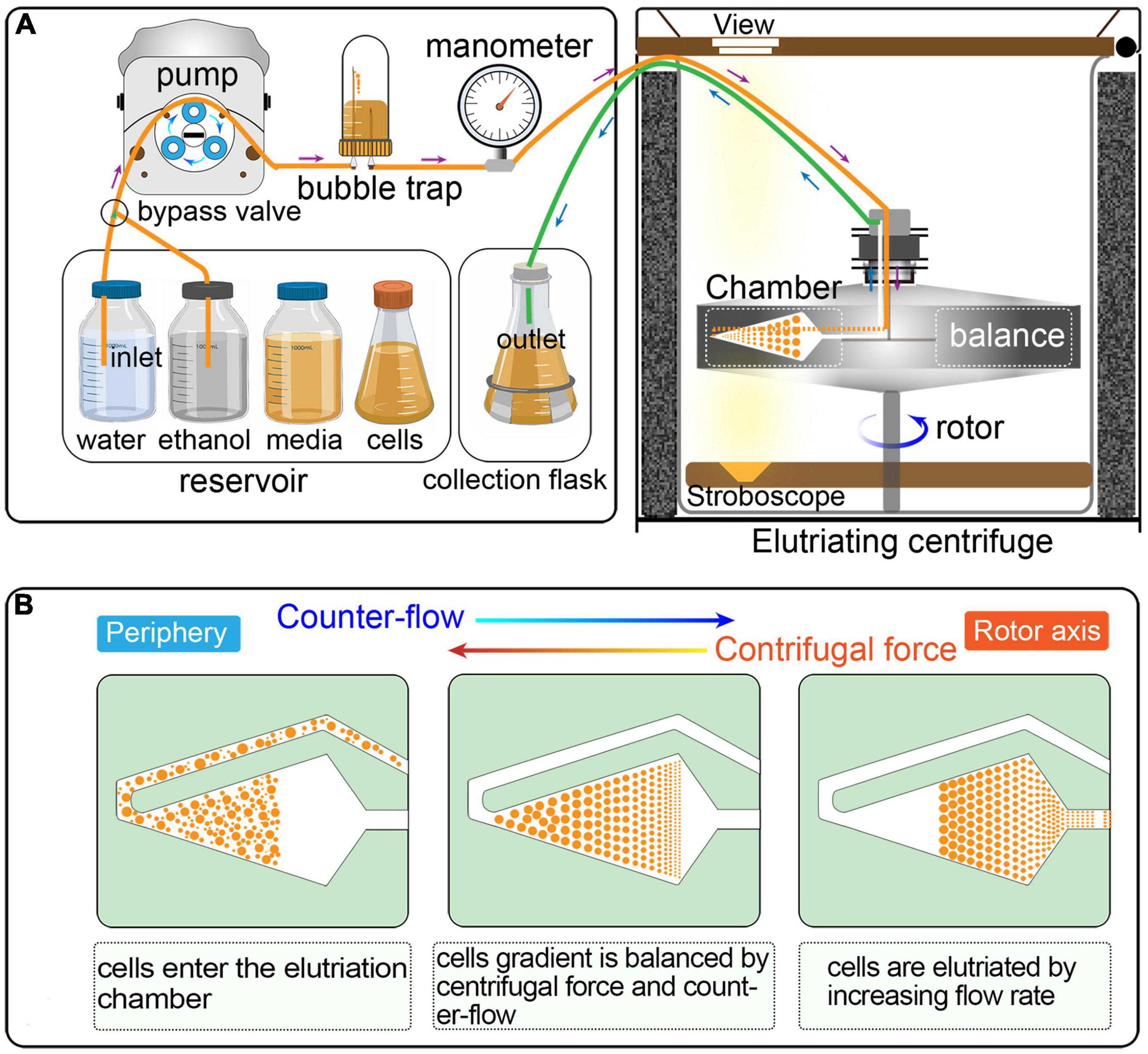
Frontiers An Optimized And Versatile Counter Flow Centrifugal Elutriation Workflow To Obtain Synchronized Eukaryotic Cells Cell And Developmental Biology

Degradation Of The Separase Cleaved Rec8 A Meiotic Cohesin Subunit By The N End Rule Pathway Journal Of Biological Chemistry

Age Related Differences In The Translational Landscape Of Mammalian Oocytes Llano 2020 Aging Cell Wiley Online Library

Meiosis Cell Division Meiosis Model How To Make Meiosis Cell Division Model Meiotic Division Youtube

Functional Interaction Between P90rsk2 And Emi1 Contributes To The Metaphase Arrest Of Mouse Oocytes Abstract Europe Pmc

Evolution Of Crossover Interference Enables Stable Autopolyploidy By Ensuring Pairwise Partner Connections In Arabidopsis Arenosa Sciencedirect
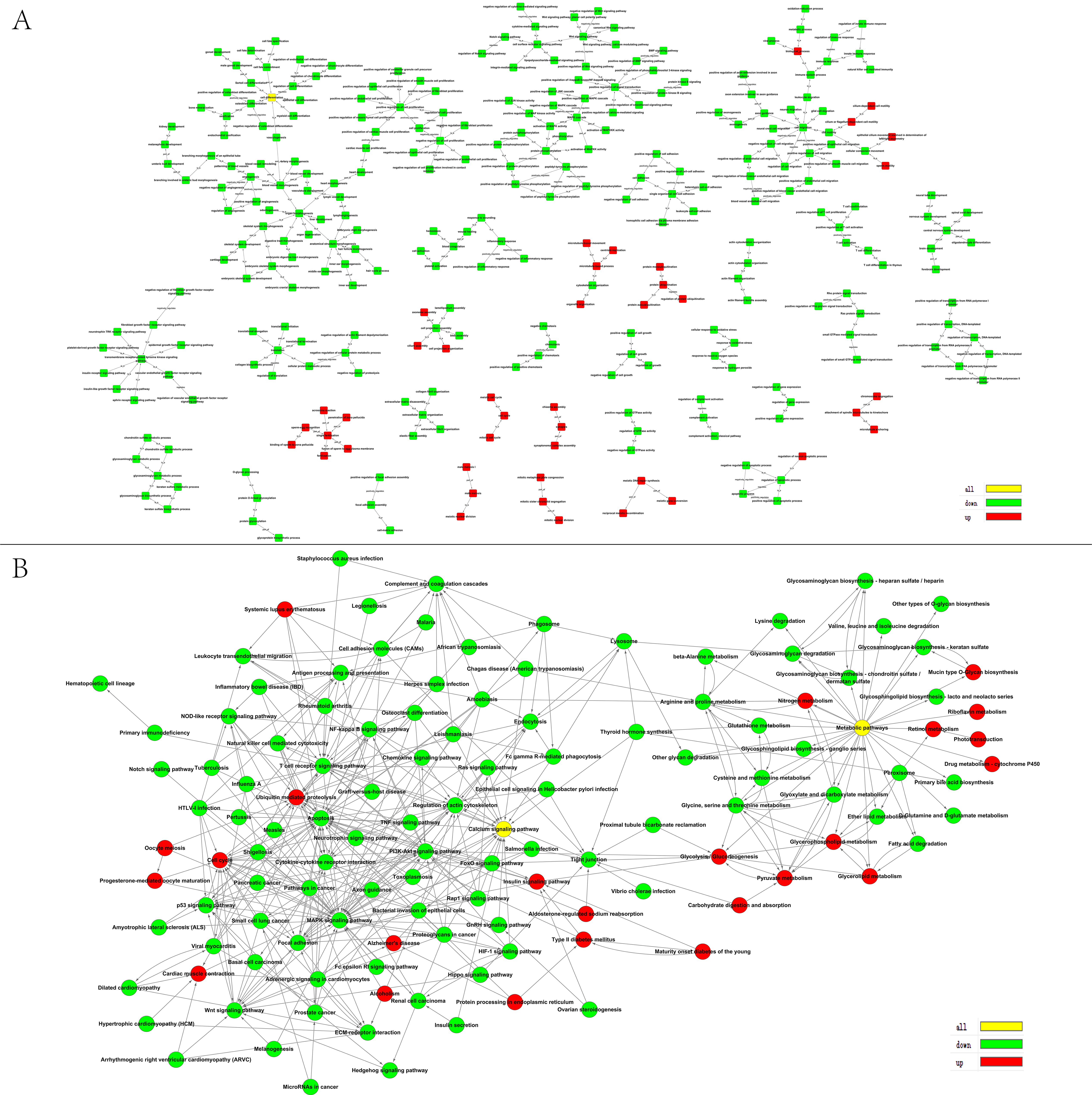
Animals Free Full Text Comprehensive Analysis Of Mirnas And Target Mrnas Between Immature And Mature Testis Tissue In Chinese Red Steppes Cattle Html









0 Response to "41 meiotic division beads diagram"
Post a Comment