37 drag the labels onto the diagram to identify the bone markings.
Image: Drag the labels onto the diagram to identify structural features associated with skeletal ... The highlighted bones articulate with which two bones? Drag the labels onto the diagram to identify structural features associated with the extrinsic muscles that move the foot and toes. To allow for flexion, the _____ unlocks the knee joint. popliteus. The tibialis posterior muscle originates at which three locations?
Art-labeling Activity: Bone Markings, Part 1. Learning Goal: To learn the bone markings. Label the bone markings. Part A. Drag the labels onto the diagram to identify the bone markings. ANSWER: Correct. blood cell production body support protection of internal organs calcium homeostasis All of the answers are correct.

Drag the labels onto the diagram to identify the bone markings.
The surface features of bones vary considerably, depending on the function and location in the body. Table 7.2 describes the bone markings, which are illustrated in (Figure 7.2.1).There are three general classes of bone markings: (1) articulations, (2) projections, and (3) holes. Transcribed image text: Art-labeling Activity: Bone Markings, Part 1 Learning Goal: To learn the bone markings Label the bone markings. Part A Drag the labels onto the diagram to identify the bone markings. Help Reset Openings Ramus Chamber within a- bone, normally filled with air Meatus Rounded passageway for blood vessels and/or nerves Fissure Elevations and Projections Projection or bump ... Correct Art-labeling Activity: Bone Markings, Part 3 Learning Goal: To learn the bone markings. Label the bone markings. Part A Drag the labels onto the diagram to identify the bone markings. ANSWER: Help Reset Fossa Tubercle Sulcus Trochanter Tuberosity Crest Line Spine
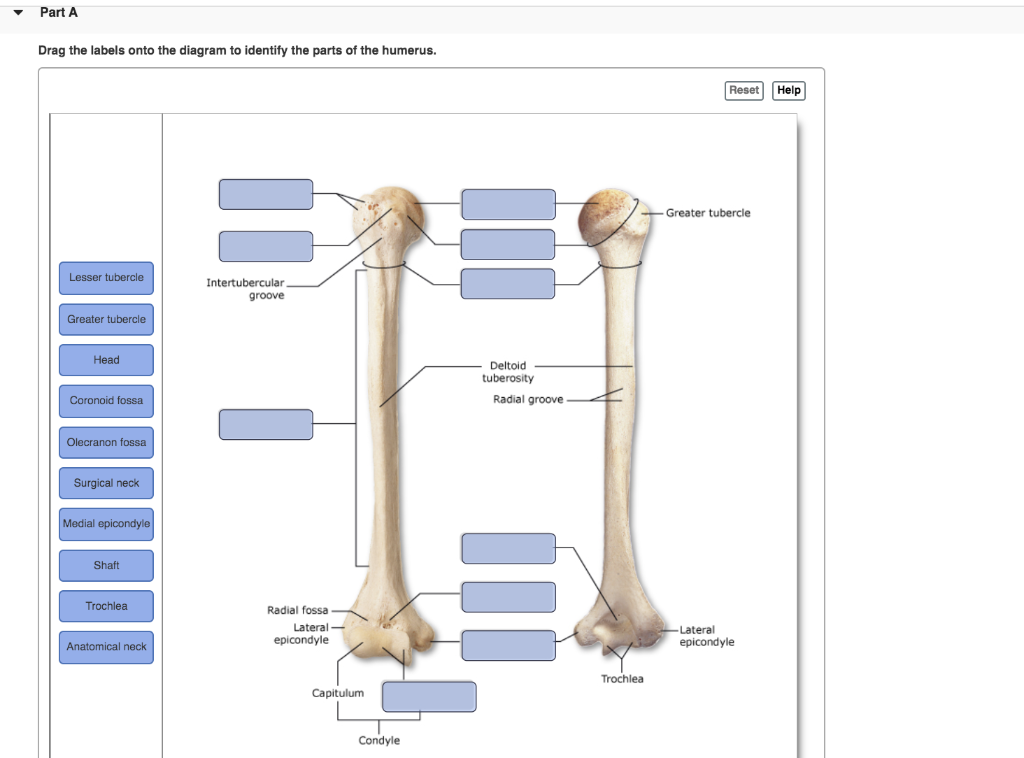
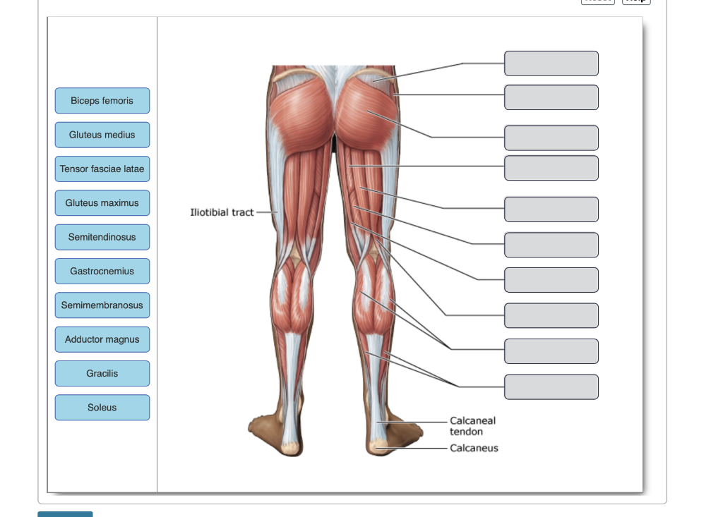
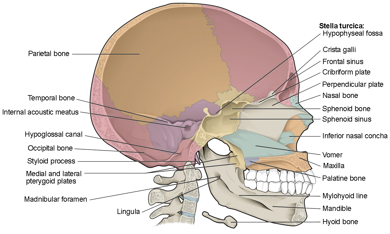
Drag the labels onto the diagram to identify the bone markings.. Anatomy and Physiology questions and answers. Identify the structures of a long bone. Part A Drag the labels onto the diagram to identify the structures. Reset Help articular cartilage articular cartilage epiphysis epiphysis medullary cavity medullary cavity epiphyseal line epiphyseal line compact bone compact bone spongy bone spongy bone ... Part A Drag the labels onto the diagram to identify the parts of the humerus. Label the bone markings on the femur. Part A Drag the labels onto the diagram. Drag the labels onto the diagram to identify the bones and markings of the skull. Image: Drag the labels onto the diagram to identify the bones and markings of ... Drag the labels onto the diagram to identify the structures found in compact bone. Osteons Circumferential lamellae. Perforating fibers. Trabeculae of spongy ...
5. Hypodermis. 6. cutaneous plexus. 7. fat. Drag the labels onto the diagram to identify the components of the integumentary system. sweat gland, hair shaft, nerve fibers, pore of sweat gland duct, tactile corpuscle, arrector pili muscle. sebaceous gland, sweat gland duct, hair follicle, lamellar corpuscle. 1. hair shaft. read through this material, identify each bone on an in-tact and/or Beauchene skull (see Figure 9.10). Note: Important bone markings are listed in the tables for the bones on which they appear, and each bone name is colored to correspond to the bone color in the figures. The Cranium The cranium may be divided into two major areas for study— Transcribed image text: Drag the labels onto the diagram to identify the bones and markings of the pelvic girdle. Reset Help postenor superior liac spine ... Answered step-by-step. If you can help me answer this I would really appreciate it. Image transcription text. Drag the labels onto the diagram to identify the structures. Reset Help ligamentum arteriosum left pulmonary arteries left subclavian artery ascending aorta left pulmonary veins. brachiocephalic trunk superior vena cava aortic arch left ...
Transcribed image text: Liauer un UUne akngs Part A Drag the labels onto the diagram to identify the bone markings. Help Reset Processes formed where ... Transcribed image text: Art-labeling Activity: Bones of the Appendicular Skeleton, Part 2 Part A Drag the labels onto the diagram to identify the bones of ... Bone Markings. The surface features of bones vary considerably, depending on the function and location in the body. Table 1 describes the bone markings, which are illustrated in (Figure 4). There are three general classes of bone markings: (1) articulations, (2) projections, and (3) holes. Correct Art-labeling Activity: Bone Markings, Part 3 Learning Goal: To learn the bone markings. Label the bone markings. Part A Drag the labels onto the diagram to identify the bone markings. ANSWER: Help Reset Fossa Tubercle Sulcus Trochanter Tuberosity Crest Line Spine
Transcribed image text: Art-labeling Activity: Bone Markings, Part 1 Learning Goal: To learn the bone markings Label the bone markings. Part A Drag the labels onto the diagram to identify the bone markings. Help Reset Openings Ramus Chamber within a- bone, normally filled with air Meatus Rounded passageway for blood vessels and/or nerves Fissure Elevations and Projections Projection or bump ...
The surface features of bones vary considerably, depending on the function and location in the body. Table 7.2 describes the bone markings, which are illustrated in (Figure 7.2.1).There are three general classes of bone markings: (1) articulations, (2) projections, and (3) holes.

Untitled (Painting) (1953/54) // Mark Rothko (Marcus Rothkowitz) American, born Russia (Latvia), 1903–1970

Untitled (Purple, White, and Red) (1953) // Mark Rothko (Marcus Rothkowitz) American, born Russia (Latvia), 1903–1970
Treatment Approaches and Options Michael G. Kaiser Regis W. Haid Christopher I. Shaffrey Michael G. Fehlings Editors














0 Response to "37 drag the labels onto the diagram to identify the bone markings."
Post a Comment