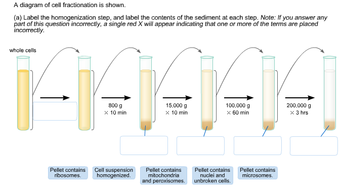
40 a diagram of cell fractionation is shown
However, as shown in Figure 2.10a, both mixing and fractionation (in this case, denitrification) can produce curves (Mariotti et al., 1988), although both relations can look linear for small ranges of concentrations. However, the equations describing mixing and fractionation processes are different and under favorable conditions, the process responsible for the curve can be identified. This is ... 23/09/2021 · Genes that pass the threshold are shown, with genes that favor cell growth in blue and those that inhibit cell growth in orange. (C) Gene Ontology of ERα-bound transcripts that favor cancer cell growth was analyzed by Database for Annotation, Visualization and Integrated Discovery (DAVID) and is presented utilizing Cytoscape software.
by B Alberts · 2002 · Cited by 20 — Analysis of protein samples by SDS polyacrylamide-gel electrophoresis. The photograph shows a Coomassie-stained gel that has been used to detect the proteins ...

A diagram of cell fractionation is shown
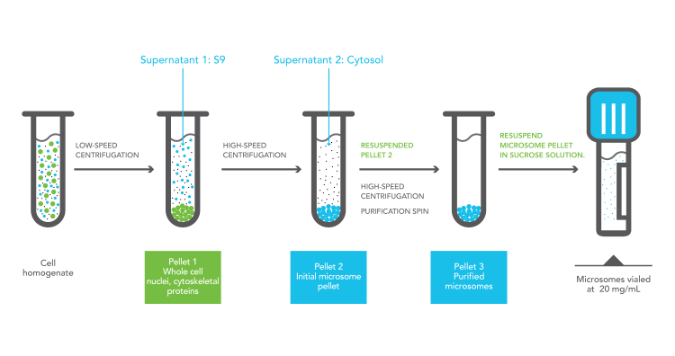
by M Lavoie · 2009 · Cited by 36 — been shown to be intimately linked to their assimilation efficien- ... Differential centrifugation diagram for subcellular fractionation of. 29/12/2015 · Cell Fractionation and RNA-Seq. Fractionation of nuclear and cytoplasmic liver RNAs was performed according to Menet et al. (2012), except for minor modifications (Supplemental Experimental Procedures). Fractionation of nuclear and cytoplasmic RNAs from MIN6 cell line (passage 30) is described in the Supplemental Experimental Procedures. RNA ... Cell fractionation is a procedure for rupturing cells, separation and suspension of cell constituents in isotonic medium in order to study their structure, ...
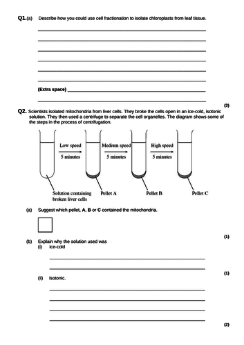
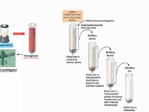
A diagram of cell fractionation is shown. The diagram shows a cell from the pancreas. (b) The cytoplasm at F contains amino acids. These amino acids are used to make proteins which are secreted from the cell. Place the appropriate letters in the correct order to show the passage of an amino acid from the cytoplasm at F until it is secreted from the cell as a protein at K. F K 11/07/2019 · Live-cell biotinylation was performed for 1 min with BP and H 2 O 2 in HEK cells stably expressing the indicated APEX2 fusion protein. APEX2 expression was visualized by anti-V5/FLAG staining (green). Biotinylated species were visualized by staining with neutravidin-AlexaFluor 647 (red). Scale bars, 10 μm. Right: pixel intensity plot of the dashed line shown in images on the left. (C) … Homogenization is a crucial step of cell fractionation. Ideally, it must ... attributed to a given subcellular component, further studies do not neces-.100 pages The process is pretty simple; you take some cells, throw them in a blender, and then centrifuge them to separate the organelles, as shown in this figure. Cell ...
09/06/2011 · Shown are five actin molecules labeled –2 to +2. The run of the protein chain is shown as a secondary structure cartoon color-coded from blue (N terminus) to red (C terminus). Interacting side chains are shown as sticks. Panels b, d, and f are stereo pairs. Panels a and b show the main longitudinal interface between molecules 0 and 2. Panels c and d show the transverse interaction … Cell suspensions are placed at first in a flow cell fitted with a device to form a liquid jet. Cells will travel through the center of a liquid jet at the rate of 5-10 m/second. Cells are then passed through the area of intense light. If the cells contain some fluorochromes, fluorescence emission can be detected using lens, beam splitters and photomultiplier tubes (Fig. 8.5). This is a method that was originally used to demonstrate the cellular location of various biochemical processes. Other uses of subcellular fractionation is to ... d. cell fractionation to study the function of specific organelles e. transmission electron microscopy (TEM) to study the movement of organelles within a living cell . d. cell fractionation to study the function of specific organelles. Which of the following clues would tell you if a cell is prokaryotic or eukaryotic? a. whether or not the cell carries out cellular metabolism b. whether or not ...
Figure 1 shows a schematic diagram of the general methods for synapse and synaptosome preparations. Synaptosomes can be further homogenized and centrifuged ... Transcribed image text: A diagram of cell fractionation is shown (a) Label the homogenization step, and label the contents of the sediment at each step, ... Many researches have shown that femtosecond laser pulsing brings these benefits to LA-ICP-MS analysis: Non-thermal mass ablation such as Coulomb explosion and direct chemical bond breaking that represents the true chemistry of the sample and is conducive in reducing the elemental/isotopic fractionation. 28/10/2021 · A. Simplified flow diagram showing the processing strategies used to determine structures of the bovine DMT with and without bound ODAs. Processing starts with extracting particles every 8 nm along the long axis of the DMT. The 8-nm particles are sorted by two-dimensional classification, with selected class averages shown, before three-dimensional refinement to generate a map of the 8-nm ...
Schematic Diagram Showing Different Steps Of Cell Fractionation Study Download Scientific Diagram
Cell fractionation is a procedure for rupturing cells, separation and suspension of cell constituents in isotonic medium in order to study their structure, ...
Ijms Special Issue Molecular Research In Radiobiology
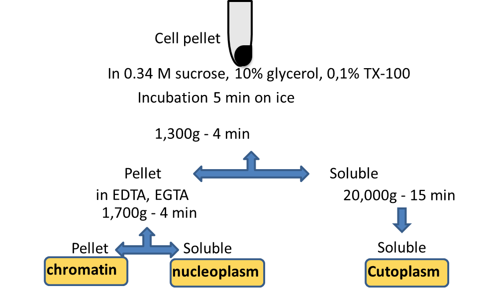
29/12/2015 · Cell Fractionation and RNA-Seq. Fractionation of nuclear and cytoplasmic liver RNAs was performed according to Menet et al. (2012), except for minor modifications (Supplemental Experimental Procedures). Fractionation of nuclear and cytoplasmic RNAs from MIN6 cell line (passage 30) is described in the Supplemental Experimental Procedures. RNA ...

A Schematic Of The Basic Cell Fractionation Presented Here Download Scientific Diagram
by M Lavoie · 2009 · Cited by 36 — been shown to be intimately linked to their assimilation efficien- ... Differential centrifugation diagram for subcellular fractionation of.
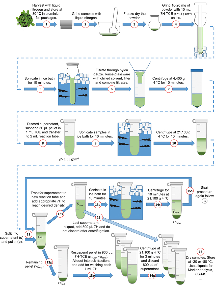
Frontiers A Benchtop Fractionation Procedure For Subcellular Analysis Of The Plant Metabolome Plant Science

Cell Fractionation An Overview Sciencedirect Topics
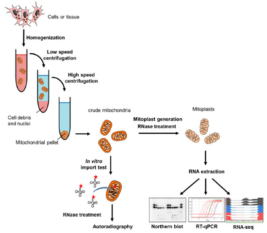
Cells Free Full Text Import Of Non Coding Rnas Into Human Mitochondria A Critical Review And Emerging Approaches Html
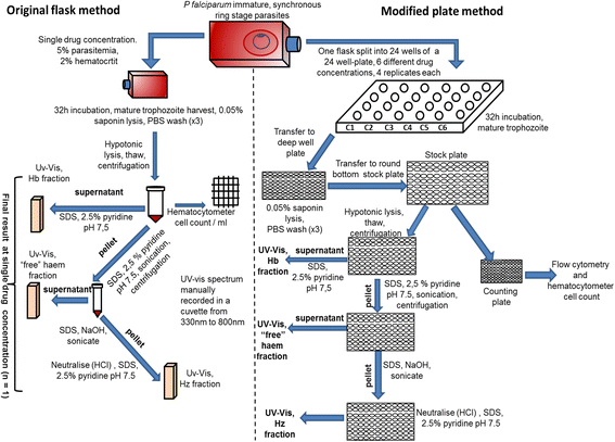
Optimization Of A Multi Well Colorimetric Assay To Determine Haem Species In Plasmodium Falciparum In The Presence Of Anti Malarials Malaria Journal Full Text
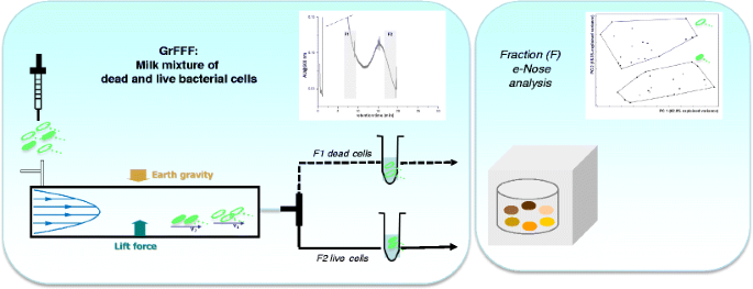
A New Analytical Platform Based On Field Flow Fractionation And Olfactory Sensor To Improve The Detection Of Viable And Non Viable Bacteria In Food Springerlink

Experiment 1 Cellbiologyolm
1
Cell Fractionation And Ultracentrifugation As Level Teaching Resources
Solved A Diagram Of Cell Fractionation Is Shown A Label Chegg Com

Cell Fractionation Definition Steps Methods Video Lesson Transcript Study Com
Cell Fractionation

Cell Fractionation And Organelle Isolation Thermo Fisher Scientific Sa

Cell Fractionation Definition Steps Methods Video Lesson Transcript Study Com

B For Biology Isolation Of Cell Organelles Subcellular Fractionation

Cell Fractionation An Overview Sciencedirect Topics

Subcellular Fractionation Of Human Liver Reveals Limits In Global Proteomic Quantification From Isolated Fractions Sciencedirect

Cell Fractionation Of U937 Cells In The Absence Of High Speed Centrifugation Protocol

Cell Fractionation
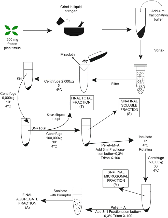
Detection And Quantification Of Protein Aggregates In Plants Springerlink

Cell Fractionation Definition Steps Methods Video Lesson Transcript Study Com

Mutant Mut Binding Site Genepharma Company Bioz Ratings For Life Science Research
Subcellular Fractions Bioivt
Cell Com
Biology Lecture No 2 11th Gw Atd On Vimeo

Fractionation An Overview Sciencedirect Topics
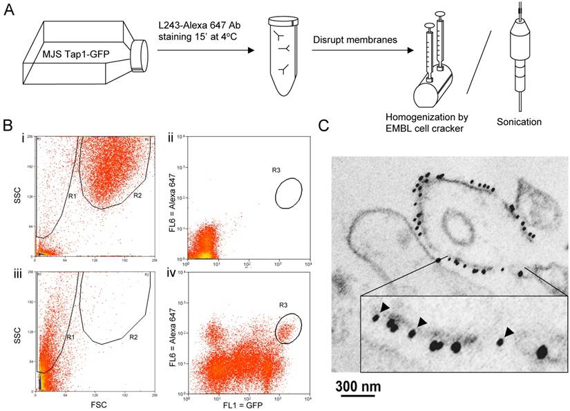
Mechanical Forces Used For Cell Fractionation Can Create Hybrid Membrane Vesicles
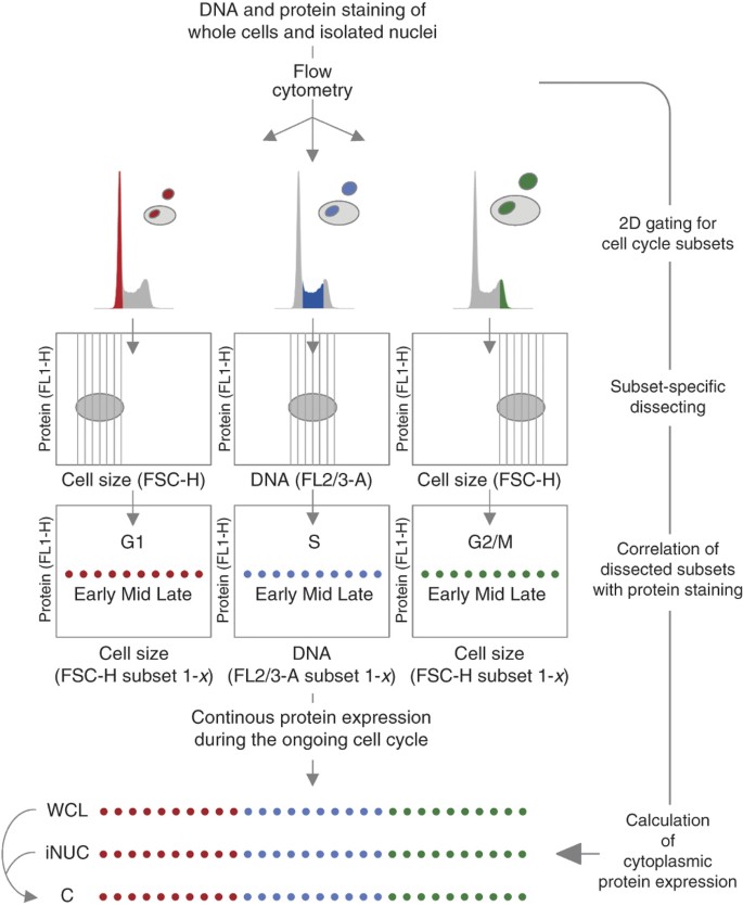
Merging High Quality Biochemical Fractionation With A Refined Flow Cytometry Approach To Monitor Nucleocytoplasmic Protein Expression Throughout The Unperturbed Mammalian Cell Cycle Nature Protocols

1 5 How We Know The Functions Of Cellular Organelles And Structures Cell Fractionation Biology Libretexts
Cell Fractionation Mp4 Youtube
Subcellular Fractionation

Subcellular Fractionation
Plos One Proteome Analysis Of Phytomonas Serpens A Phytoparasite Of Medical Interest

Sequential Fractionation And Isolation Of Subcellular Proteins From Tissue Or Cultured Cells Sciencedirect

1 Schematic Separation Of Organelles By Differential Centrifugation Download Scientific Diagram
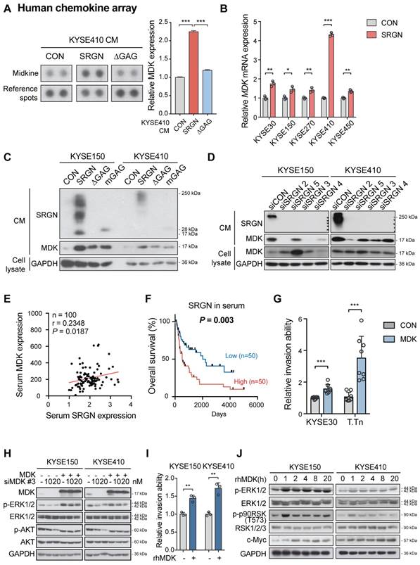
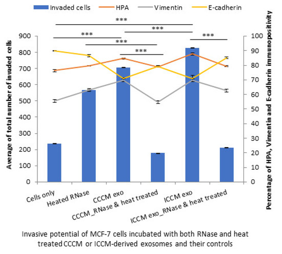
Significance Of Serglycin And Its Binding Partners In Autocrine Promotion Of Metastasis In Esophageal Cancer

Cell Fractionation Kit High Throughput Ht Ab109718 Abcam

Cell Fractionation 512 Words Studymode
0 Response to "40 a diagram of cell fractionation is shown"
Post a Comment