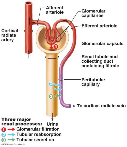
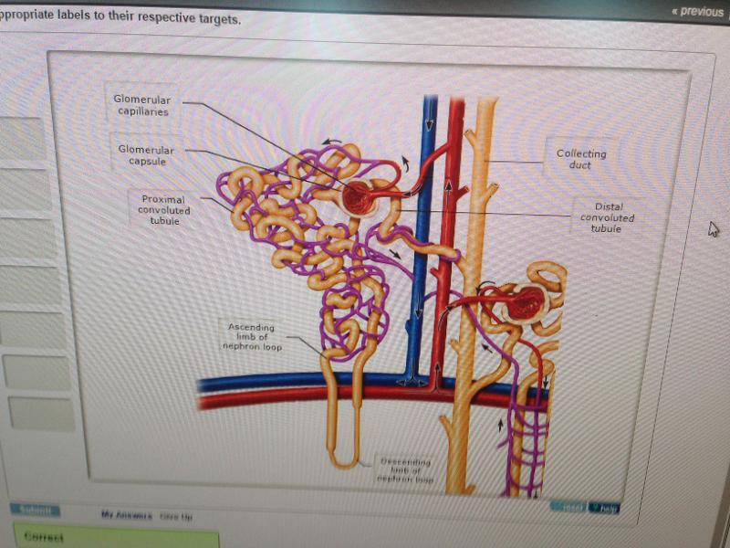
38 drag the labels onto the diagram to identify the blood vessels of the kidneys.
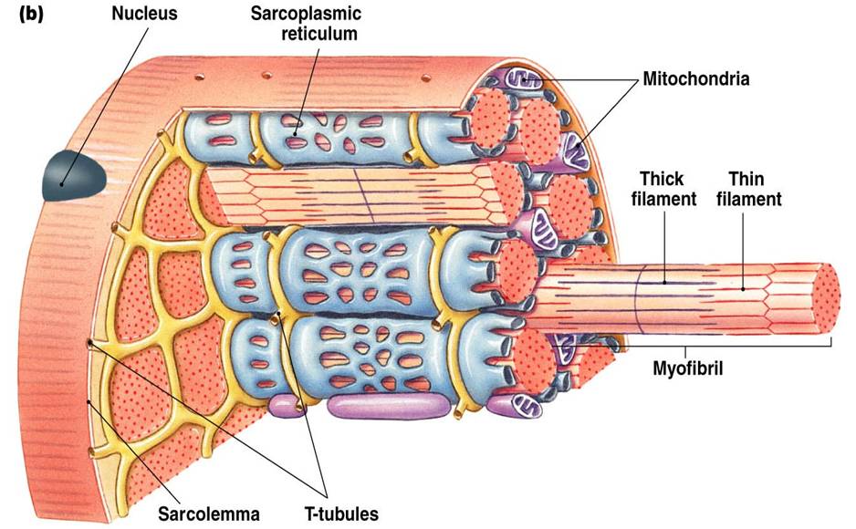
The tunica intima , or inner-most layer of blood vessels, is made of endothelial tissue. Art-labeling Activity: Figure 32.1 (1 of 2) Label the major types of blood vessels and their layers. Part A Drag the labels onto the diagram to identify the major types of blood vessels and their layers. Home 10 2 skeletal muscle anatomy and physiology drag the labels onto the diagram to identify structural features associated with skeletal muscle. 20 1 structure and function of blood vessels anatomy and physiology. Drag the labels onto the diagram to identify the steps in a reaction both with and without enzymes.
From the left. On this diagram show how the steps in the formation of aldosterone. Part a drag the labels onto the diagram to identify features of cell signaling and receptors. Drag each one into its correct location in the table. Many factors including blood ph and blood pressure and producing erythrocytes.
Drag the labels onto the diagram to identify the blood vessels of the kidneys.
Identify the vessels through which blood travels within the pulmonary circuit, beginning from the right ventricle of the heart and ending at the left atrium Create a flow chart showing the major systemic arteries through which blood travels from the aorta and its major branches, to the most significant arteries feeding into the right and left ... This diagram shows the different ions and chemicals that are secreted and reabsorbed along the nephron. solved part a identifying the structures the kidney plete the diagram below using the following steps place the pink labels which indicate interstitial fluid osmolarity in mosm l onto the correct pink tar s note that the numbers inside the ... Drag the appropriate labels to their respective targets. Study ap chapter 6 bones and skeletal tissues flashcards taken from chapter 6 of the book human anatomy physiology. Name each chamber and provide the name and general route of its associated great vessels. Drag the labels onto the diagram to identify the surface anatomy of the heart.
Drag the labels onto the diagram to identify the blood vessels of the kidneys.. This is due to its rich blood supply—it houses 90-95% of the kidney's blood vessels. At specific points, extensions of the renal cortex called renal columns pass through the renal medulla to-ward the renal pelvis. The renal columns house blood vessels Figure 24.3 Internal anatomy of the kidney, including the nephron. Renal hilum Renal pelvis -kidneys filter blood - urine travels thru ureter - stored in urinary bladder ... Drag the labels to the diagram to identify each organ. kidneys. ureters. bladder. urethra-renal cortex = outer shell around the medulla. renal artery and vein = blood delivery-renal medulla = center of the kidney. renal pyramids ... renal vessels. major calyx ... Drag The Labels Onto The Diagram To Identify Structures And Students can also color the image to identify the major structures of the nephron. Label the major structures of the nephron and associated structures. This is the currently selected item. General overview of the raas system. There are about 1000000 nephrons in each human kidney. Drag the labels onto the diagram to identify the parts of the kidney. ... Chronic and acute renal failure impairs all of the functions carried out by the kidneys and, as a consequence, the functions of most other body systems. ... The collection of blood vessels supplying the protective connective tissue layers surrounding the brain may ...
View the full answer. Transcribed image text: Drag the labels onto the diagram to identify the blood vessels of the kidneys. Reset Help Renal vein Renal artery Glomerulus Interlobar artery Afferent arteriole Efferent arteriole Segmental artery Peritubular capillaries Interlobar vein Cortical radiate artery Cortical radiate vein Arcuate vein ... peritoneal cavity; and the lining of all blood vessels. t Glandular epithelium forms most of the body glands. Epithelia occur at the boundary between two different envi-ronments. The epidermis of the skin, for example, lies between the inside and the outside of the body. Most substances that enter into the body or are released from the body ... Drag the labels onto the diagram to identify the structures of the upper respiratory system. Note the direct link of the renal arteries and renal veins into the aorta and vena cava respectively. Complete the labeling of the diagram to correctly identify the urinary system organs. Drag the labels onto the diagram to identify the stem cells and stages of white blood cell and pl. Drag the labels onto the diagram to identify the stages in which the lagging strand is synthesized.. Part a nucleotide pairing drag the labels onto the diagram to identify how nucleotides pair up.
- secretions are released into blood stream - exocrine glands - cells that retain their connection with the epithelial surface - duct is present - secretion released onto surface of epithelium -types of exocrine glands 1. Halocrine - cell dies and releases content - sebaceous gland 2. Drag the appropriate labels to their respective targets. Drag the labels onto the diagram to identify the blood types that correspond to specific blood typing test results. Drag the appropriate labels to their respective targets. Study ap chapter 5 the integumentary system flashcards taken from chapter 5 of the book human anatomy physiology. Drag the labels onto the diagram to identify the effects of isotonic hypotonic and hypertonic solutions on red blood cells. Drag the labels onto the diagram to identify respiratory system structures. Lungs are to the respiratory system as the liver is to the system. Label the structures of the lower respiratory tract. Classifying epithelia identify the types of epithelia. The layers do not contain any blood vessels and one surface of the cells lines the cavity of the organ. Show transcribed image text drag the correct labels onto the diagram to identify the structures and molecules involved in translation. Solved drag the labels onto the diagram to identity ...
1
Show transcribed image text drag the labels onto the diagram to identify structural features associated with skeletal muscle. Label this diagram. Answer Correct Chapter Test Chapter 10 Question 4 Endomysium Numbers to the left identify the spinal nerves and indicate where the nerve roots leave the vertebral canal.
Cardiovascular System Circulatory System Parts And Functions
Drag the labels onto the diagram to identify the origins of the cranial nerves (VII - XII). look at pic The accumulation of blood during an epidural or subdural hemorrhage creates debilitating pressure on the brain and, without help, death is imminent.
Associate Degree Nursing Physiology Review
Drag the labels onto the diagram to identify the stem cells and stages of white blood cell and platelet production. Drag the labels onto the diagram to identify structural features associated with skeletal muscle. After each piece of the lagging stand is complete it is released from dna polymerase. Ap chapter 5 the integumentary system.
1
Label the arteries of the abdomen. Drag and drop the text labels onto the boxes next to the heart diagram. The arteries are the blood vessels that deliver oxygen rich blood from the heart to the tissues of the body. These are arteries near the body surface at which you can feel a pulse. The coronary arteries is the main artery that taking blood ...

A P2 Lab 13 Hw A P2 Lab 12 Hw A P2 Lab 11 Hw A P2 Lab 10 Hw Lab 9 Hw Lab 8 Hw A P2 Lab 1 Hw A P2 Lab 2 Hw A P2 Lab
Drag The Labels Onto The Diagram To Identify The Structures And Ligaments Of The Shoulder Joint - Anatomy And Physiology Fluids And Transport The Cardiovascular System Blood Vessels And Circulation Viva Open : Movement, target, target motion direction, prime mover, origin .. Joint stability is provided instead by the rotator cuff muscles ...
Web As Uky Edu
Labeled Diagram of the Human Kidney. The human kidneys house millions of tiny filtration units called nephrons, which enable our body to retain the vital nutrients, and excrete the unwanted or excess molecules as well as metabolic wastes from the body. ... The blood supply to all these structures occurs through the branches and sub-branches of ...
Narrowed Small Blood Vessels Linked To Fatigue In Me Cfs Health Rising
Drag and drop the text labels onto the boxes next to the heart diagram It consists of two periods: one during which the heart muscle relaxes and refills with blood, called diastole, following a period of robust contraction and pumping of blood, dubbed systole Chu, H April 5, 2021 9:30 AM - 5:00 PM ET Register Now!

Clinical Evaluation Of Autonomic Disorders Sciencedirect
Show transcribed image text drag the labels onto the diagram to identify structural features associated with skeletal muscle. 20 1 structure and function of blood vessels anatomy and physiology drag the labels onto the diagram to identify structural features associated with skeletal muscle. 5163 x 2217 pixel.
Lamission Edu
Drag the labels to their appropriate locations on the diagram below. Label diagram 20 3 drag the labels onto the diagram to identify the superficial features of the heart on the posterior surface. The kidneys of terrestrial mammals conserve water in the body by concentrating urine.
Anatomy And Physiology Fluids And Transport The Cardiovascular System Blood Vessels And Circulation Viva Open
Signal recognition particle SRP binds to the signal peptide as it emerges from the ribosome. part a drag the labels onto the diagram to identify the part a drag the labels onto the diagram to identify the stages of the life cycle not all labels will be used answer chapter 8 reading quiz question 2. Proteins all begin their synthesis in the ...

Labeling Activity 1 V Able Bluedoor Llc V All Good 5 Homeworklib
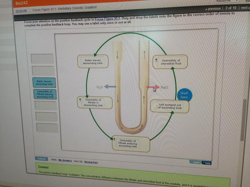
Drag the labels onto the diagram to identify the events resulting from decreasing ECF volume, and the body's homeostatic responses. (Note: If two labels can be equally placed in two targets the labels should be placed in alphabetical order from top to bottom.)
Print Final Exam Flashcards Easy Notecards
Drag the appropriate labels to their respective targets. Study ap chapter 6 bones and skeletal tissues flashcards taken from chapter 6 of the book human anatomy physiology. Name each chamber and provide the name and general route of its associated great vessels. Drag the labels onto the diagram to identify the surface anatomy of the heart.

Jaypeedigital Ebook Reader
This diagram shows the different ions and chemicals that are secreted and reabsorbed along the nephron. solved part a identifying the structures the kidney plete the diagram below using the following steps place the pink labels which indicate interstitial fluid osmolarity in mosm l onto the correct pink tar s note that the numbers inside the ...

Chapter 47 Sensory Perception Bio 140 Human Biology I Textbook Libguides At Hostos Community College Library
Identify the vessels through which blood travels within the pulmonary circuit, beginning from the right ventricle of the heart and ending at the left atrium Create a flow chart showing the major systemic arteries through which blood travels from the aorta and its major branches, to the most significant arteries feeding into the right and left ...
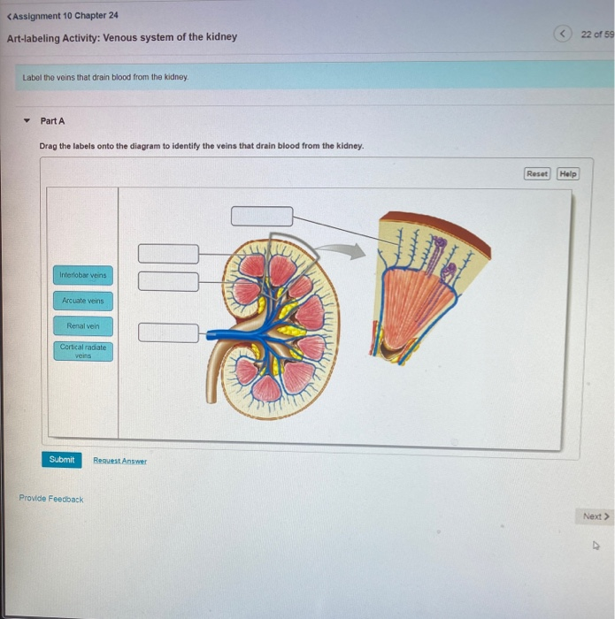
Solved Assignment 10 Chapter 24 Art Labeling Activity Chegg Com
Pearsonhighered Com
Hrdcnepal Org
Chapter 25 The Urinary System Mastering Flashcards Easy Notecards
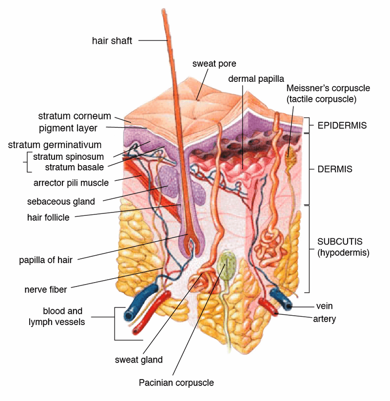
Integumentary System Definition Organs Functions Diseases

Chapter 25 Flashcards Quizlet

Structure And Function Of Blood Vessels Anatomy And Physiology
Etd Ohiolink Edu
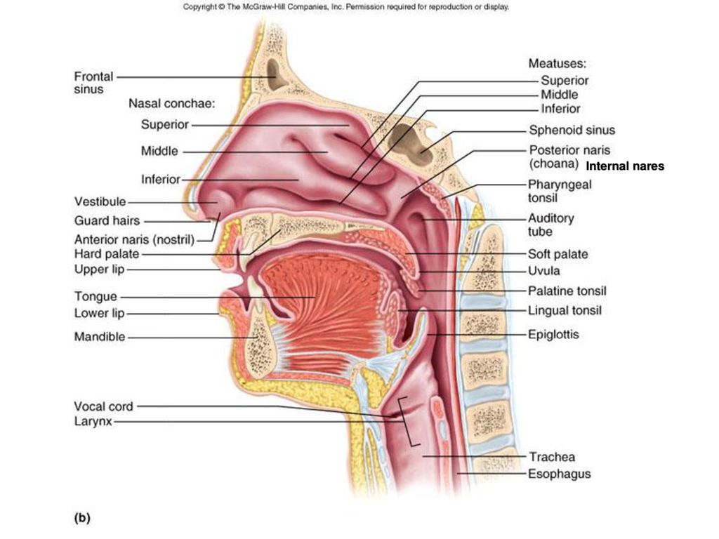
The Respiratory System Ppt Download
Integumentary System Definition Organs Functions Diseases
Chapter 25 The Urinary System Mastering Flashcards Easy Notecards

A P2 Lab 13 Hw A P2 Lab 12 Hw A P2 Lab 11 Hw A P2 Lab 10 Hw Lab 9 Hw Lab 8 Hw A P2 Lab 1 Hw A P2 Lab 2 Hw A P2 Lab

Peterson Michael Book Images

Human Anatomy And Physiology Posts Facebook

Homeostatic Regulation Of The Vascular System Anatomy And Physiology Ii

File 2101 Blood Flow Through The Heart Jpg Wikimedia Commons

A P2 Lab 13 Hw A P2 Lab 12 Hw A P2 Lab 11 Hw A P2 Lab 10 Hw Lab 9 Hw Lab 8 Hw A P2 Lab 1 Hw A P2 Lab 2 Hw A P2 Lab

A P2 Lab 13 Hw A P2 Lab 12 Hw A P2 Lab 11 Hw A P2 Lab 10 Hw Lab 9 Hw Lab 8 Hw A P2 Lab 1 Hw A P2 Lab 2 Hw A P2 Lab
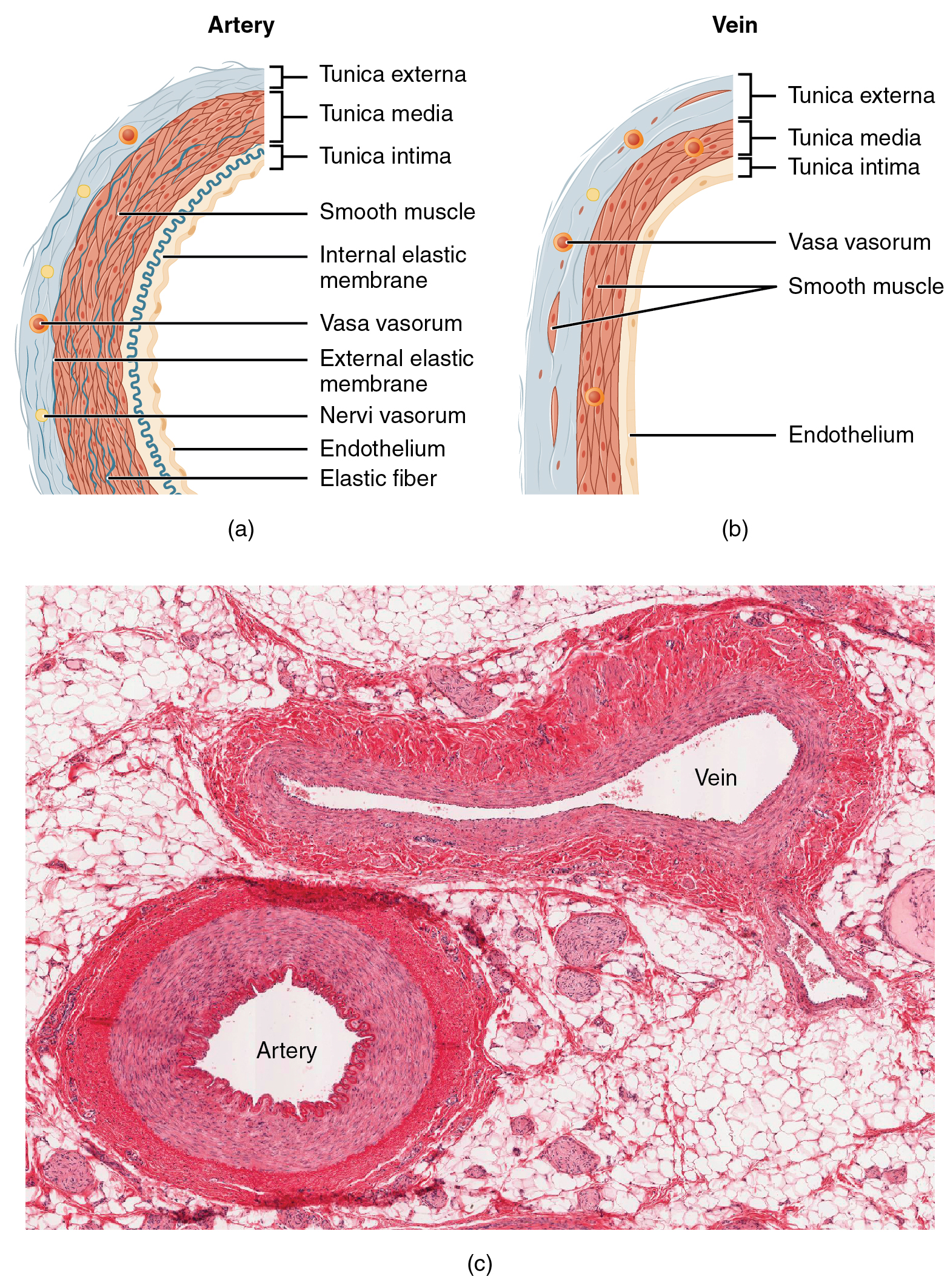
Structure And Function Of Blood Vessels Anatomy And Physiology
Etd Ohiolink Edu

Ch 25 Hw Pdf Ch25hw Ch25hw Due 11 59pmonsunday April26 2015 Gradingpolicy 1 Parta Course Hero
Electrolyte Fluid Balance

A P2 Lab 13 Hw A P2 Lab 12 Hw A P2 Lab 11 Hw A P2 Lab 10 Hw Lab 9 Hw Lab 8 Hw A P2 Lab 1 Hw A P2 Lab 2 Hw A P2 Lab

60 Brain Stuff For Sciencing Ideas Anatomy And Physiology Physiology Human Anatomy And Physiology
0 Response to "38 drag the labels onto the diagram to identify the blood vessels of the kidneys."
Post a Comment