42 diagram of a sarcomere
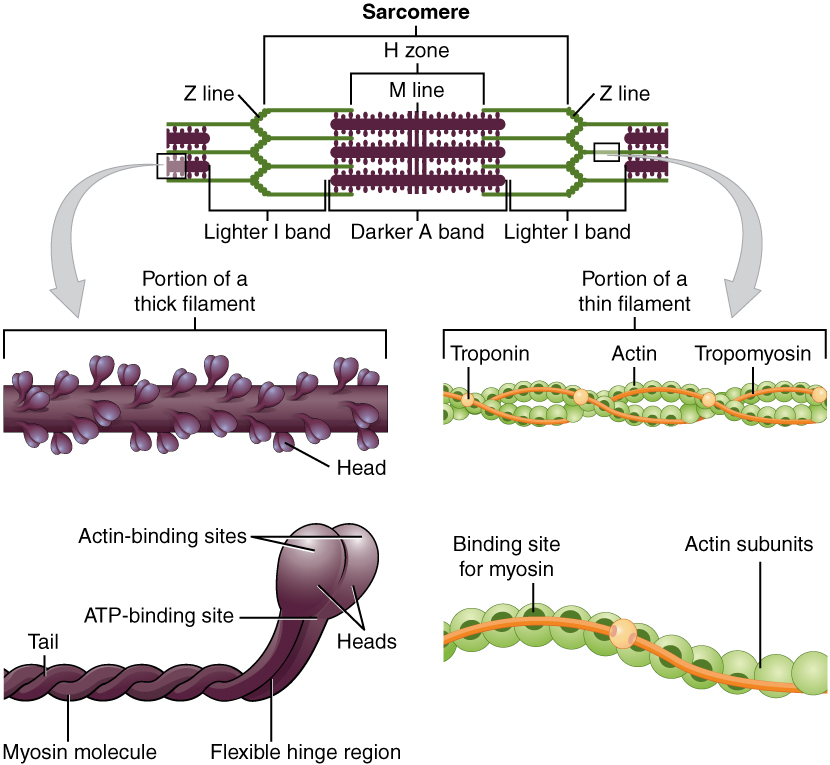
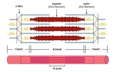
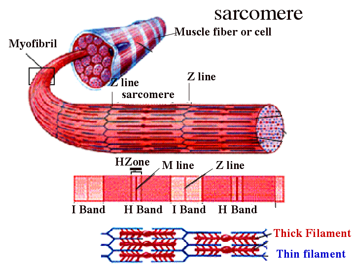
Jan 23, 2019 · Diagram Of Sarcomere. A sarcomere is the basic unit of striated muscle tissue. It is the repeating unit between two Z lines. Skeletal muscles are composed of tubular muscle cells which. sarcomere. Schematic: The Z line is depicted in black, myosin in red, actin in green/gray, and tropomyosin in blue. Image: MPI of Molecular Plant Physiology. Each myofibril is made up of contractile sarcomeres AND Drawing labelled diagrams of the structure of a sarcomere.
This site was designed for students of anatomy and physiology. It contains textbook resources, such as chapter review guides, homework sets, tutorials, and printable images. Each chapter has a practice quiz and study tips for learning the topic.

Diagram of a sarcomere
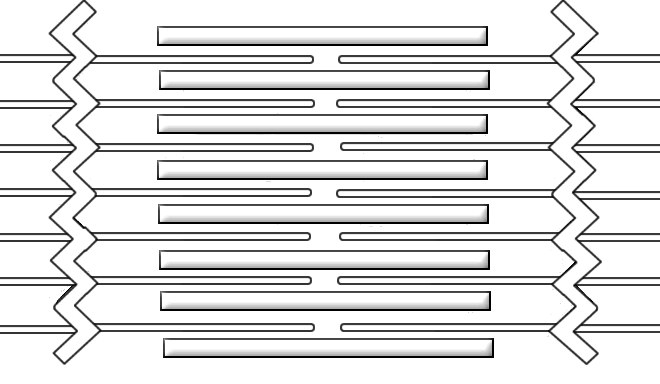
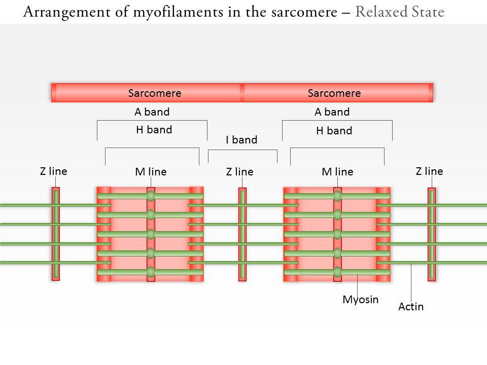
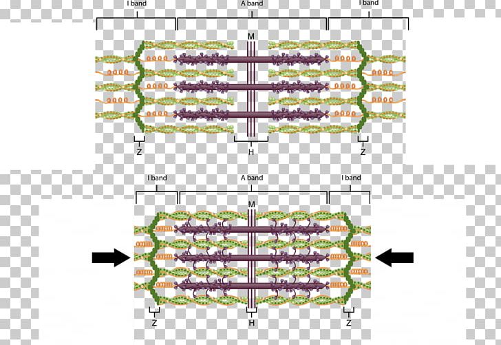
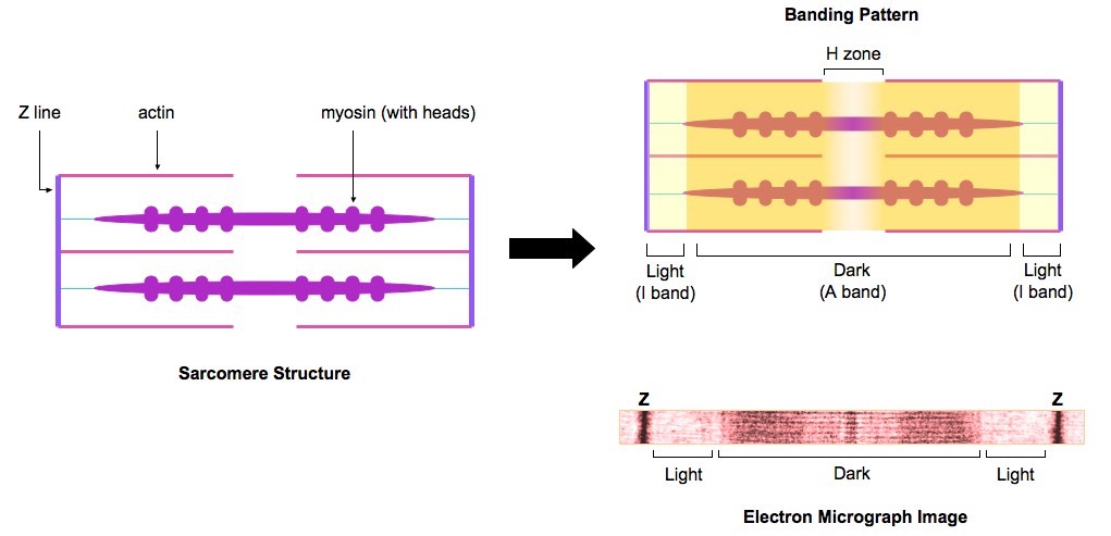
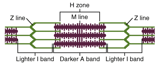
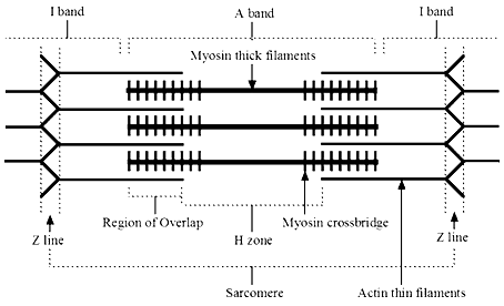
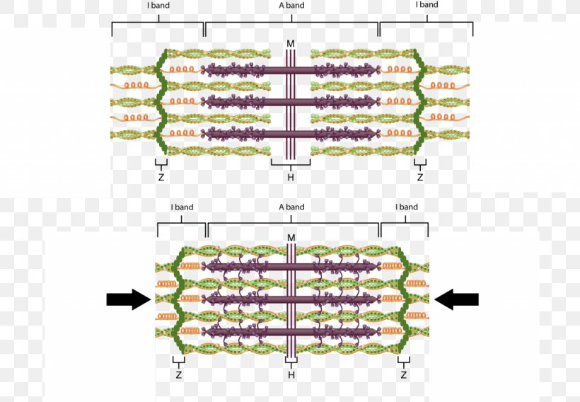
The diagram above shows part a myofibril called a sarcomere. This is the smallest unit of skeletal muscle that can contract. Sarcomeres repeat themselves over and over along the length of the myofibril. Here is a quick reminder of all the muscle structures involved: sarcomere. within myofibrils are organized consecutively composed of 2 filaments (actin and myosin) actin. thin myofilament has troponin and tropomyosin has active site that the myosin heads bind to. myosin. thick myofilament band, and a sarcomcre, When you have finished, draw a contracted sarcomere in the space beneath the figure and label the same structures, as well as the light and dark bands Z disc Myosin Actin filaments Figure 6—3 1. Looking at vour diagram of a contracted sarcomere from a slightly different angle, which region of the sarcomere shortetvs
Diagram of a sarcomere. May 13, 2019 · Sarcomere, a component in the structure of muscle and/or the attachment of Above: Diagram of the unit within a muscle cell that is known as a sarcomere. A sarcomere is the basic unit of striated muscle tissue. It is the repeating unit between two Z lines. Skeletal muscles are composed of tubular muscle cells which. Jan 23, 2019 · A sarcomere is the basic unit of striated muscle tissue. It is the repeating unit between two Z lines. Skeletal muscles are composed of tubular muscle cells which. Sarcomeres are composed of thick filaments and thin filaments. The thin filaments Look at the diagram above and realize what happens as a muscle contracts. 7.1.2022 · Active Immunity Definition. What is Active Immunity? Active Immunity occurs when your body becomes immune to a disease due to a vaccine, a successful infection in the past, or exposure to what will become an infection in the future.. The condition of being immune to a particular disease by having had it or being inoculated against it. Titin / ˈ t aɪ t ɪ n /, also known as connectin, is a protein that in humans is encoded by the TTN gene. Titin is a giant protein (contraction for Titan protein), greater than 1 µm in length, that functions as a molecular spring which is responsible for the passive elasticity of muscle.It comprises 244 individually folded protein domains connected by unstructured peptide sequences.
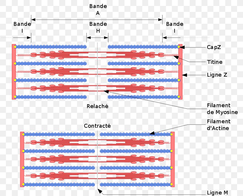
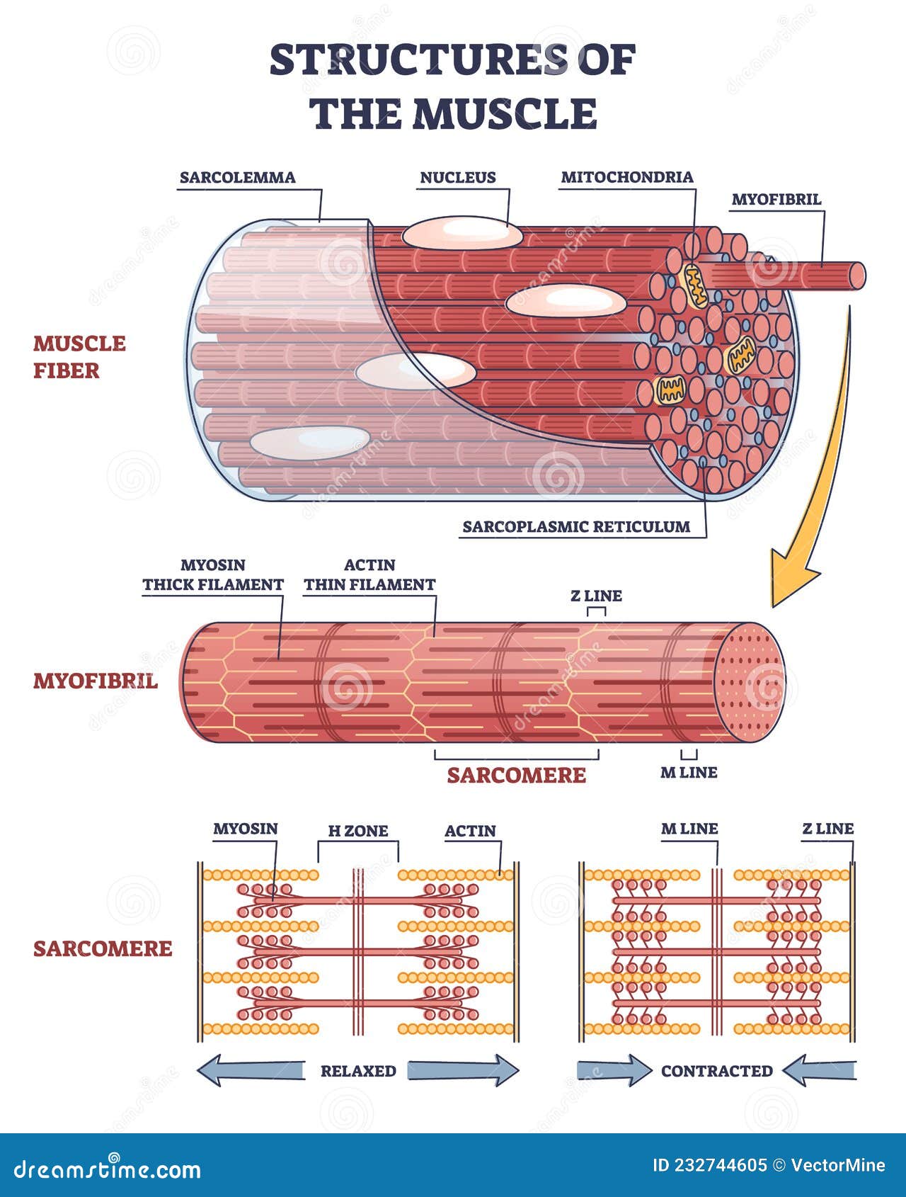
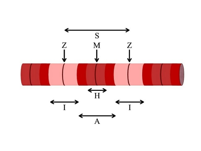
Sarcomere structure When viewed under a microscope, muscle fibers of varied lengths are organized in a stacked pattern. The myofibril strands, thereby actin and myosin, form bundles of filament arranged parallel to one another. When a muscle in our body contracts, it is understood that the way this happens follows the sliding filament theory. Start studying Sarcomere Diagram. Learn vocabulary, terms, and more with flashcards, games, and other study tools. A sarcomere describes as the distance between two Z discs or Z lines. When a muscle contracts in our body the distance reduces between the Z discs. The central region of the A zone (H zone), contains only thick filaments (myosin), and became short during contraction. (b) Schematic diagram of a cardiac sarcomere. The sarcomere is the fundamental unit of contraction and is defined as the region between two Z-lines. Each sarcomere consists of a central A-band (thick filaments) and two halves of the I-band (thin filaments). The I-band from two adjacent sarcomeres meets at the Z-line.
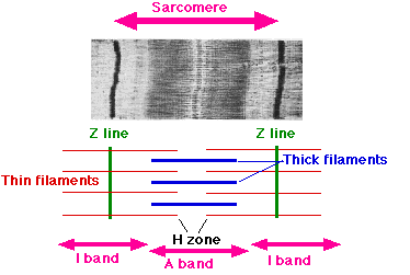
Diagram and micrograph of a sarcomere The I band is that part of the sarcomere that contains thin filaments, while the A band contains an area of overlap between the thin and the thick filaments. Mass Haul Diagram Explained. Whirlpool Duet Dryer Parts Diagram. Minecraft Circle Diagram. Standing Rigging Diagram. 3 Position Switch Wiring Diagram. sarcomere is the ultimate source of muscle power, the mechanical output of muscle depends ... If we want to introduce the Hill model as a block diagram, the Figure 8 will show it well. Figure8: Hill- type muscle model – block diagram. Muscle model Contractile component (CC) … (b) Schematic diagram of a cardiac sarcomere. The sarcomere is the fundamental unit of contraction and is defined as the region between two Z-lines. Each sarcomere consists of a central A-band (thick filaments) and two halves of the I-band (thin filaments). The I-band from two adjacent sarcomeres meets at the Z-line. 4.5.2021 · Cardiac muscle is similar to skeletal muscle in that it is striated and that the sarcomere is the contractile unit, contraction being achieved by the relationship between calcium, troponins and the myofilaments. This article will consider the structure of cardiac muscle as well as relevant clinical conditions.
Sarcomere diagram. 14 terms. MaddieSheedlo97. Sarcomere/ Sarcoplasmic Reticulum/ T-Tubule. 46 terms. Rayeanna_Hoff. Other sets by this creator. Lecture 4 - Mass Balances. 24 terms. dethomas1. Environmental Legislation (1&2) 87 terms. dethomas1. Renewable energy technology mid term. 13 terms. dethomas1. Environmental Management Exam.
Sarcomere Structure. To understand the sliding filament model requires an understanding of sarcomere structure. A sarcomere is defined as the segment between two neighbouring, parallel Z-lines. Z lines are composed of a mixture of actin myofilaments and molecules of the highly elastic protein titin crosslinked by alpha-actinin.
Start studying Sarcomere. Learn vocabulary, terms, and more with flashcards, games, and other study tools.
Click here to get an answer to your question ✍️ Draw the diagram of a sarcomere of skeletal muscle showing different regions.
band, and a sarcomcre, When you have finished, draw a contracted sarcomere in the space beneath the figure and label the same structures, as well as the light and dark bands Z disc Myosin Actin filaments Figure 6—3 1. Looking at vour diagram of a contracted sarcomere from a slightly different angle, which region of the sarcomere shortetvs
sarcomere. within myofibrils are organized consecutively composed of 2 filaments (actin and myosin) actin. thin myofilament has troponin and tropomyosin has active site that the myosin heads bind to. myosin. thick myofilament
The diagram above shows part a myofibril called a sarcomere. This is the smallest unit of skeletal muscle that can contract. Sarcomeres repeat themselves over and over along the length of the myofibril. Here is a quick reminder of all the muscle structures involved:













0 Response to "42 diagram of a sarcomere"
Post a Comment