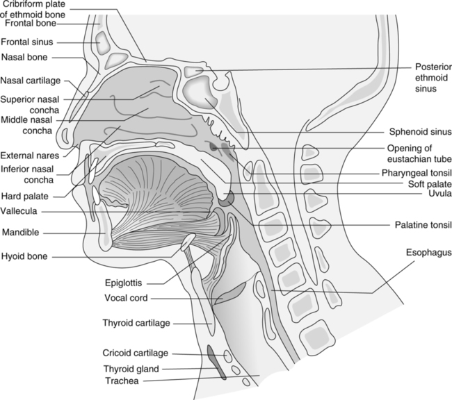
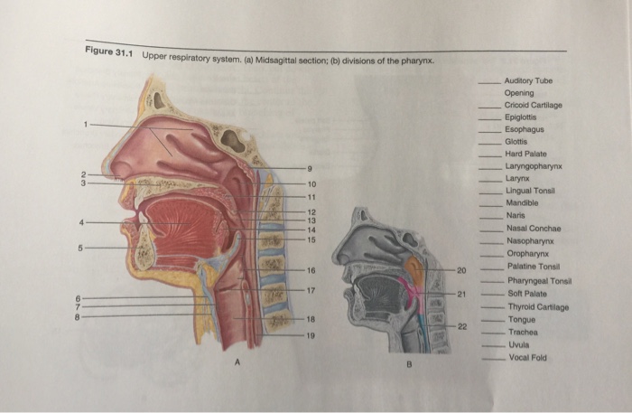
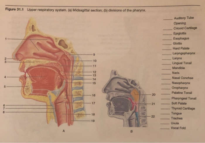
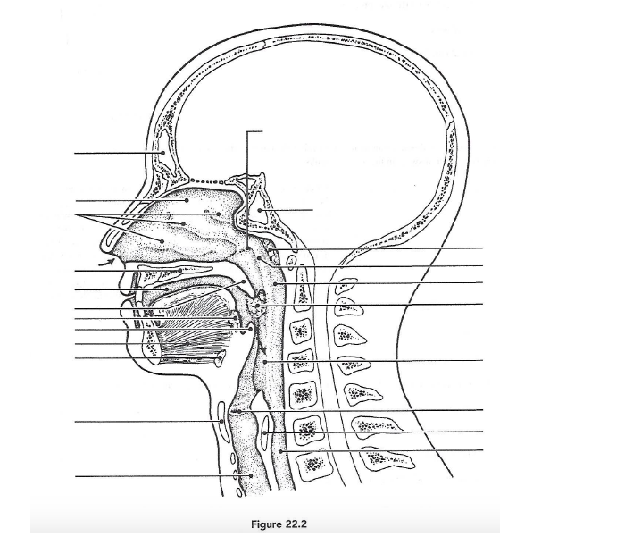
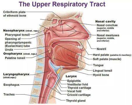
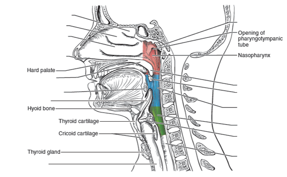
40 complete the labeling of the diagram of the upper respiratory structures (sagittal section)
...Upper and lower Respiratory System Structures 1. Complete the labeling of the model of the respiratory structures (sagittal section) shown below. the diagram of the upper respiratory structures (sagittal section) Opening of pharyngotympanic Hard palate Hyoid bone Thyroid... upper respiratory cartilage and ligament… 28 terms. laura_lovlie. Labeled Sagittal Section of the Upper Re…
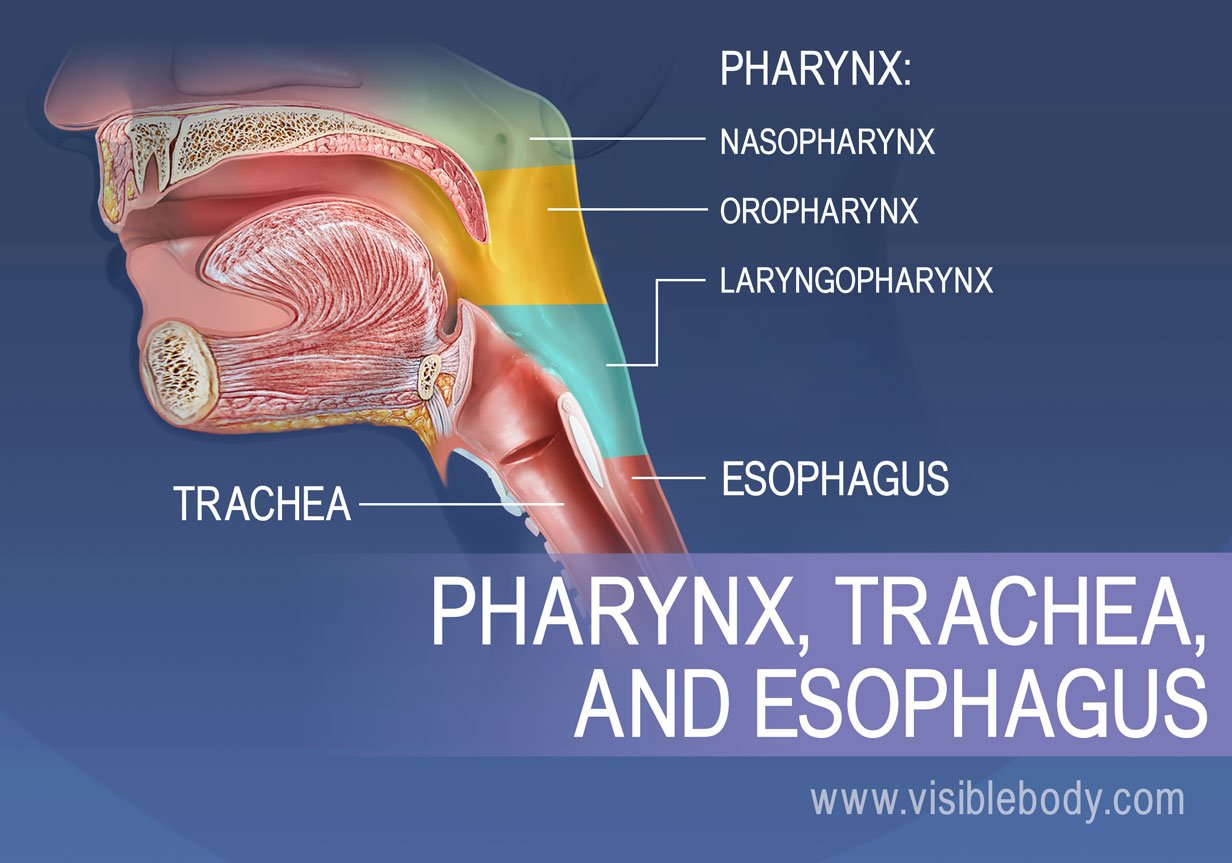
Human respiratory system, the system in humans that takes up oxygen and expels carbon dioxide. The major organs of the respiratory system include the nose, pharynx, larynx, trachea Overview mechanism and anatomy of the respiratory tract; passaging air from the mouth and nose to the lungs.

Complete the labeling of the diagram of the upper respiratory structures (sagittal section)
1. Complete the labeling of the diagram of the upper respiratory structures (sagittal section). Cribriform plate of ethmoid bone. The more common site for lodging of a foreign object that has entered the respiratory passageways Согтестлу label all structures provided with leader lines on... Jan 20, 2022 · Complete the labeling of the diagram of the upper respiratory structures (sagittal section). complete the labeling of the diagram of the upper respiratory structures sagittal section frontal sinus cribriform plate of ethmoid bone superior, the nose is an olfactory and respiratory organ it consists of nasal skeleton which houses the nasal ... Label the parts of the upper respiratory system: cente, epiglottis, ... Fill in the blanks with the parts of the conducting zone in the correct sequence: ...
Complete the labeling of the diagram of the upper respiratory structures (sagittal section). Describe how the respiratory system processes oxygen and CO2; Compare and contrast the functions of upper respiratory tract with the lower respiratory tract. Figure 12.1 image description: This diagram shows the location of the heart in the thorax (sagittal and anterior views). The sagittal view labels read (from top, clockwise): first rib, aortic arch, thoracic arch, esophagus, inferior vena cava, diaphragm, thymus, trachea. The anterior view lables read (from top, clockwise): mediastinum, arch of aorta, pulmonary trunk, left auricle, left … Diagram of the Human Respiratory System (Infographic). An exchange of oxygen and carbon dioxide takes place in the alveoli, small structures within the lungs. The carbon dioxide, a waste gas, is exhaled and the cycle begins again with the next breath. An upper respiratory tract infection is any infection that involves the nasal cavity, paranasal sinuses, pharynx, or larynx, and it's most often caused by an invading pathogen like a virus. These droplets can then land in the mouths or noses of people nearby, or get inhaled into the upper airways.
Browse our listings to find jobs in Germany for expats, including jobs for English speakers or those in your native language. ...of the upper respiratory structures (sagittal section) quizlet. In the diagram at left which of the labeled structures is the thoracic duct. Ap ii review sheet 36 anatomy Name lab timedate anatomy of the respiratory system upper and lower respiratory system structures 1. Due to respiratory... Complete the labeling of the diagram of the upper respiratory structures (sagittal section). Frontal sinus -.. Cribriform plate- of eth mold bone StLhPJtO 1 --- cO& Sphenoidal sinus Opening of auditory tube Nasopharynx tv4erva- Y"- -- Hard palate- - I ongue Hyoid bone- Thyroid cartilage of larynx Cricoid cartilage Thyroid gland yaLLfLcc ... Human Respiratory System. The Upper Airway and Trachea. All these structures act to funnel fresh air down from the outside world into your body. Structure The lungs are paired, cone-shaped organs which take up most of the space in our chests, along with the heart.
Upper and lower respiratory system structures. 53 if 3an c t. Anatomy of blood vessels conduction system of the heart electrocardiography human cardiovascular physiology. In the diagram at left which of the labeled structures is the thoracic duct. Two pairs of vocal folds are found in the larynx. The respiratory portion begins at the level where alveoli first appear in the final branches of the bronchioles. Now look at a sagittal section of the palate (slide 115) and compare respiratory epithelium of the nasal passage View Image to the stratified squamous epithelium of the oral cavity. ...Upper and Lower Respiratory System Structures 1. Complete the labeling of the diagram of the upper respiratory structures (sagittal section). ...RESPIRATORY SYSTEM The respiratory system consists of all the organs involved in breathing. These include the nose, pharynx, larynx... R'*.f Labrime/Da," l0 tg-lg Upper and Lower Respiratory System Structures 1. Complete the labeling of the diagram of the upper respiratory structures (sagittal section). \p iot go' P }''-'t ^r"\ hs;l Opening of pharyngotympanic tube IQ, Hyoid bone Thyroid gland \tP 2. ]1wo pairs of vocal folds are found in the larynx.
upper respiratory tract - consisting of the nose, nasal cavity and the pharynx. lungs have appearance of a glandlike structure. stage is critical for the formation of all conducting airways. At birth, fluid in the upper respiratory tract is expired and fluid in the lung aveoli is rapidly absorbed this...
Name lab timedate anatomy of the respiratory system upper and lower respiratory system structures 1. Complete the labeling of the diagram Your nose is divided into the external nose and the internal nasal cavity. Know and be able to label the following. Due to respiratory movement 50.
Upper respiratory tract: This includes the nose, mouth, and the beginning of the trachea (the The structure of the lungs includes the bronchial tree - air tubes branching off from the bronchi into Its purpose is to diagnose obstructive diseases of the respiratory system. It produces a diagram...
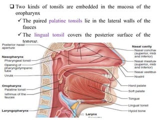
The structures of the upper respiratory system warm and clean the air by trapping particles and pollutants before they travel into the lungs. 1. The Nose and Nasal Cavities Provide Airways for Respiration. The nasal cavities are chambers of the internal nose.
Complete the labeling of the diagram of the upper respiratory structures (sagittal section). ... Appropriately label all structures provided with leader lines on the ...
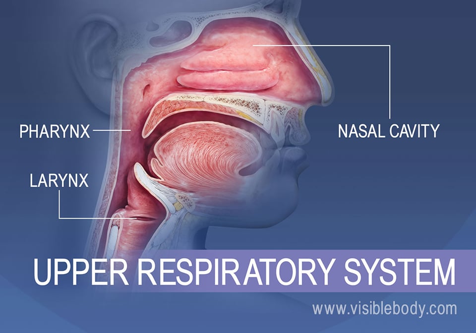
Complete respiratory system. The upper airways or upper respiratory tract includes the nose and nasal passages, paranasal sinuses, the pharynx, and the portion of the larynx above the vocal folds (cords).
06.10.2021 · The primary motor cortex (M1) is essential for voluntary fine-motor control and is functionally conserved across mammals1. Here, using high-throughput transcriptomic and epigenomic profiling of ...
What is the upper respiratory tract definition, its parts, structure, and anatomy, function of the upper respiratory system, and how it works, labeled diagram. The nostrils, the two round or oval holes below the external nose, are the primary entrance into the human respiratory system [5]. Lying just...
The respiratory system is represented by the following structures, shown in Figure 1 The epiglottis, the first piece of cartilage of the larynx, is a flexible flap that covers the glottis, the upper region of the larynx, during swallowing to . Figure 2.Anterior and sagittal sections of the larynx and the trachea.
# 1. Complete the labeling of the diagram of the upper respiratory structures (sagittal section). # 8. Appropriately label all structures provided with leader lines on the diagrams below. Produce a serous fluid that reduces friction during breathing movements and helps.
Transcribed image text : Upper and Lower Respiratory System Structures 1. Complete the labeling of the model of the respiratory structures (sagittal section) shown below.
Drag the labels onto the diagram to identify the structures of the upper respiratory system. Which pair are the true vocal cords superior or inferior. Anatomy of upper respiratory system sagittal section the game ends when you get all 30 questions correct or when you give up published.
Upper and Lower Respiratory System Structures Complete the labeling of the diagram of the upper respiratory structures (sagittal section)). Larger in diameter? More horizontal? Appropriately label all structures provided with leader lines on the diagrams below.
By the end of this section, you will be able to: ... labeling each of the major vessels. It is beyond the scope of this text to name every vessel in the body. However, we will attempt to discuss the major pathways for blood and acquaint you with the major named arteries and veins in the body. Also, please keep in mind that individual variations in circulation patterns are not uncommon. …
List the structures that make up the respiratory system. Describe how the respiratory system Upper part of trachea. The major organs of the respiratory system function primarily to provide The nares open into the nasal cavity, which is separated into left and right sections by the nasal septum.
... 36 283 Upper and Lower Respiratory System Structures 1. Complete the labeling of the diagram of the upper respiratory structures (sagittal section). 2.
BIO MEDICAL INSTRUMENTATION. Enter the email address you signed up with and we'll email you a reset link.
A&P II - Review Sheet 36 - Anatomy of the Respiratory System ... Image: Know and be able to label the following. Two pairs of vocal folds are found in the ...
We always make sure that writers follow all your instructions precisely. You can choose your academic level: high school, college/university, master's or pHD, and we will assign you a writer who can satisfactorily meet your professor's expectations.
Complete the labeling of the diagram of the upper respiratory structures (sagittal section). 9 con. Opening of pharyngotympanic tube Nasopharynx Hard palate ...
Upper and Lower Respiratory System Structures 1. Complete the labeling of the diagram of the upper respiratory structures (sagittal section). 11. What portions of the respiratory system are referred to as anatomical dead space? All but the respiratory zone structures (respiratory...
Feb 11, 2018 · It is formed by 9 supportive cartilages, intrinsic and extrinsic muscles and a mucous membrane lining. It is a short inch tube that is located in the throat, inferior to the hyoid bone and tongue and anterior to the esophagus. Complete the labeling of the diagram of the upper respiratory structures (sagittal section). 2.
EssEntials strEngth training Conditioning NatioNal StreNgth aNd CoNditioNiNg aSSoCiatioN THird EdiTion
Shark gill. Schematic section of a gill element in frontal plane from the left side of a shark, showing (A) most important anatomical structures, (B) terminology and functional units. (C) Block diagram and cross section of gill filaments. Black arrows indicate direction of water flow; white arrows, blood flow.
The respiratory system has a complex physiology and is responsible for multiple functions. There are multiple roles performed by the respiratory system: pulmonary ventilation, external respiration, internal respiration, transportation of gases and homeostatic control of respiration.
Hier sollte eine Beschreibung angezeigt werden, diese Seite lässt dies jedoch nicht zu.
LAB REPORT 31 Nam Respiratory Structures Section checklist for your study of the human model and the shore luck. GROSS DESCRIPTION: A. Received fresh labeled with patient's name, designated 'right upper lobe wedge', is an 8.0 x 3.5 x 3.0 cm wedge of lung which has an 11.5 cm...
1. Complete the labeling of the diagram of the upper respiratory structures {sagittal section). Hard palate. 8. Appropriately label all structures provided with leader lines on the diagrams below. 9. Trace a molecule of oxygen from the nostrils to the pulmonary capillaries of the lungs: Nostrils *.
Upper limb. With a labeled diagram, you can see all of the main structures of an organ system together on one page - great for helping you to memorise the appearance of several structures and their relations. Take a look at the labeled diagram of the respiratory system above.
...Respiratory System Upper and Lower Respiratory System Structures 1. Complete the labeling of the diagram of the upper respiratory structures (sagittal Your answer : b. pH will decrease and PCO2 will increase. Stop & Think Questions: Which of the following can cause respiratory acidosis?…
Complete the labeling of the diagram of the upper respiratory structures (sagittal section).SphenoidalsinusHyoid bone----->-,Thyroidcartilage ReviewSheet 365479.Trace a molecule of oxygen from the nostrils to the pulmonary capillaries of the lungs: Nostrils ~10.Match the terms in...
55 Likes, 13 Comments - UCLA VA Physiatry Residency (@uclava_pmrresidency) on Instagram: “Resident’s Corner: Name: David Huy Blumeyer, MD Year in …
Label the parts of the upper respiratory system: cente, epiglottis, ... Fill in the blanks with the parts of the conducting zone in the correct sequence: ...
Jan 20, 2022 · Complete the labeling of the diagram of the upper respiratory structures (sagittal section). complete the labeling of the diagram of the upper respiratory structures sagittal section frontal sinus cribriform plate of ethmoid bone superior, the nose is an olfactory and respiratory organ it consists of nasal skeleton which houses the nasal ...
1. Complete the labeling of the diagram of the upper respiratory structures (sagittal section). Cribriform plate of ethmoid bone. The more common site for lodging of a foreign object that has entered the respiratory passageways Согтестлу label all structures provided with leader lines on...












0 Response to "40 complete the labeling of the diagram of the upper respiratory structures (sagittal section)"
Post a Comment