38 Simple Squamous Epithelium Labeled Diagram
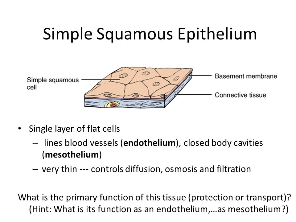
Label Simple Squamous Epithelium - epithelium slide 22 ... Here are a number of highest rated Label Simple Squamous Epithelium pictures on internet. We identified it from obedient source. Its submitted by government in the best field. We understand this nice of Label Simple Squamous Epithelium graphic could possibly be the most trending subject when we allocation it in google gain or facebook. Simple Squamous Epithelium |Inrtroducrion , Anatomy & Function Simple squamous epithelium is the tissue that creates from one layer of squamous cells which line surfaces. The squamous cells are thin, large, and flat, and consisting of around nucleus. These tissues have polarity like other epithelial cells and consist of a distinct apical surface with special membrane proteins.
Simple Squamous Epithelium - Definition and Examples ... Examples of Simple Squamous Epithelia. Simple squamous epithelia are found in a variety of locations, starting from capillaries to the alveoli of lungs, and nephrons of kidneys. Most of these cells arise from the ectoderm, or outermost layer of cells in the embryo. However, some simple squamous epithelial tissues are also derived from the mesoderm or middle layer of embryonic cells. These are called the mesothelia, especially in pathological studies.
Simple squamous epithelium labeled diagram
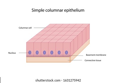
Simple Columnar Epithelium Labeled Diagram These labelled diagrams should closely follow the current Science (simple squamous epithelium). ORIGIN: columnar epithelium with goblet cells. TISSUE . Simple Squamous Epithelium Definition Simple squamous epithelium is a single sheet of cells joined together by flat cells cal. Simple columnar epithelium (40X) Primate small intestine. Epithelial Tissue: Structure with Diagram, Function, Types ... Simple squamous: Blood vessel lining, air sac lining of lungs: A single layer of flat cells having irregular boundaries: Transport by diffusion and where minimal protection is required: Simple Cuboidal: The tubular lining of kidneys, glandular ducts: A single layer of short cylindrical cells. It may have microvilli as in proximal convoluted tubules PDF Basic Histo diagrams labelled in colour - 2005 These labelled diagrams should closely follow the current Science courses in histology, anatomy and ... squamous epithelium) ORIGIN: ectoderm lamina propria ... Covers the external surface. blood vessel lined by endothelium (simple squamous epithelium) ORIGIN: mesoderm lumen GENERALISED SECTION epithelium OF THE BODY connective tissue beneath ...
Simple squamous epithelium labeled diagram. Ileum: Anatomy, histology, composition, functions | Kenhub It is made up of simple squamous epithelium and a connective tissue layer underneath (lamina propria serosae). A characteristic feature of the ileum is the Peyer's patches lying in the mucosa. It is an important part of the GALT (gut-associated lymphoid tissue). One patch is around 2 to 5 centimeters long and consists of about 300 aggregated ... Simple squamous epithelium- structure, functions, examples Simple squamous epithelium definition. Simple squamous epithelium is a type of simple epithelium that is formed by a single layer of cells on a basement membrane. It is a type of epithelium formed by a single layer of squamous or flat cells present on a thin extracellular layer, called the basement membrane. This epithelium is also termed the pavement epithelium because the cells appear like tiles on a floor when viewed from the apical surface. Simple Columnar Epithelium: A Labeled Diagram and ... Bodytomy provides a labeled diagram to help you understand the structure and function of simple columnar epithelium. Home / Uncategorized / Simple Columnar Epithelium: A Labeled Diagram and Functions. Epithelium is a tissue that lines the internal surface of the body, as well as the internal organs. Simple epithelium is one of the types of epithelium that is divided into simple columnar epithelium, simple squamous epithelium, and simple cuboidal epithelium. simple squamous epithelium diagram - Google Search ... Simple columnar epithelium with very regular line-up of nuclei. This photo displays cardiac muscle tissue. This tissue type is striated, branched, and has 1-2 nuclei. Cardiac muscle tissue can be recognized by intercalated disks (dark thickened part). Cardiac muscle cells are bigger than smooth muscle cells and smaller than skeletal muscle cells.
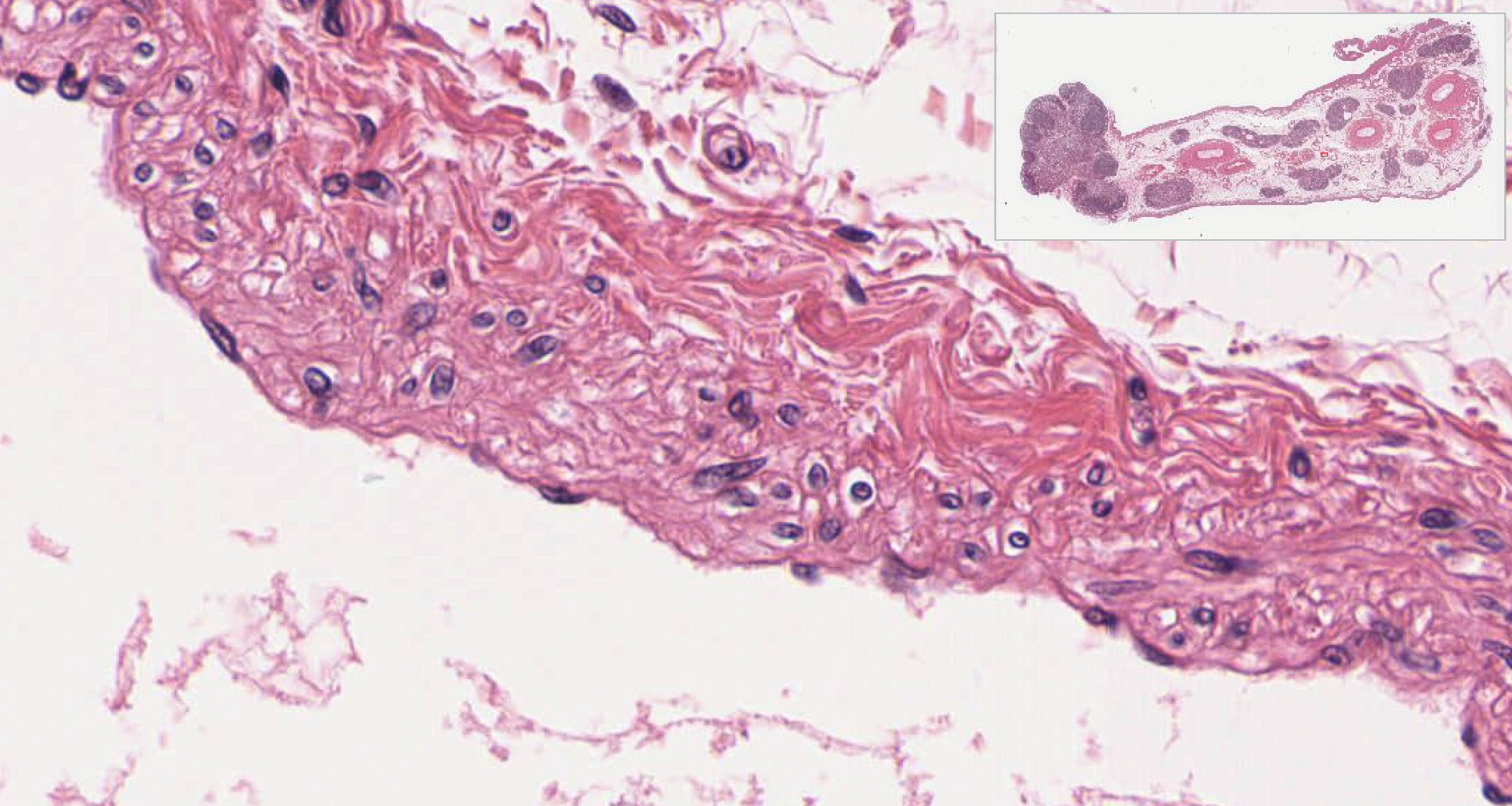
DOC Anatomy Review: Respiratory Structures 1. simple squamous epithelium. 2. alveolar macrophages. 3. surfactant-secreting cells • The wall of an alveolus is primarily composed of simple squamous epithelium, or Type I cells. Gas exchange occurs easily across this very thin epithelium. Simple squamous epithelium - Eugraph The outer wall is composed of a single layer of flat cells (a simple squamous epithelium). Two of the nuclei (stained purple) are indicated by arrows labeled n. Two areas of cytoplasm (stained pink) are indicated by arrows labeled c. The simple squamous epithelium shown here is the outer wall of the glomerular capsule. More information about glomerular capsules and related structures is available in the section on the kidney. Uterus Histology - Best Place to Learn Veterinary Anatomy ... #4. The outer perimetirum is lined by simple squamous epithelium or mesothelium. So this is the uterus slide of animal. Okay, let's find these characteristics from the uterus slide picture. Uterus histology slide with labeled diagram. Do you want to know the details histology of different layers of uterus with real slide and labeled diagram? Simple Squamous Epithelium: Location and Diagram - Video ... The simple squamous epithelium location specifically exists in the lining of the blood vessels like the arteries, veins, and capillaries. It is also found lining the alveoli or air sacs within the ...
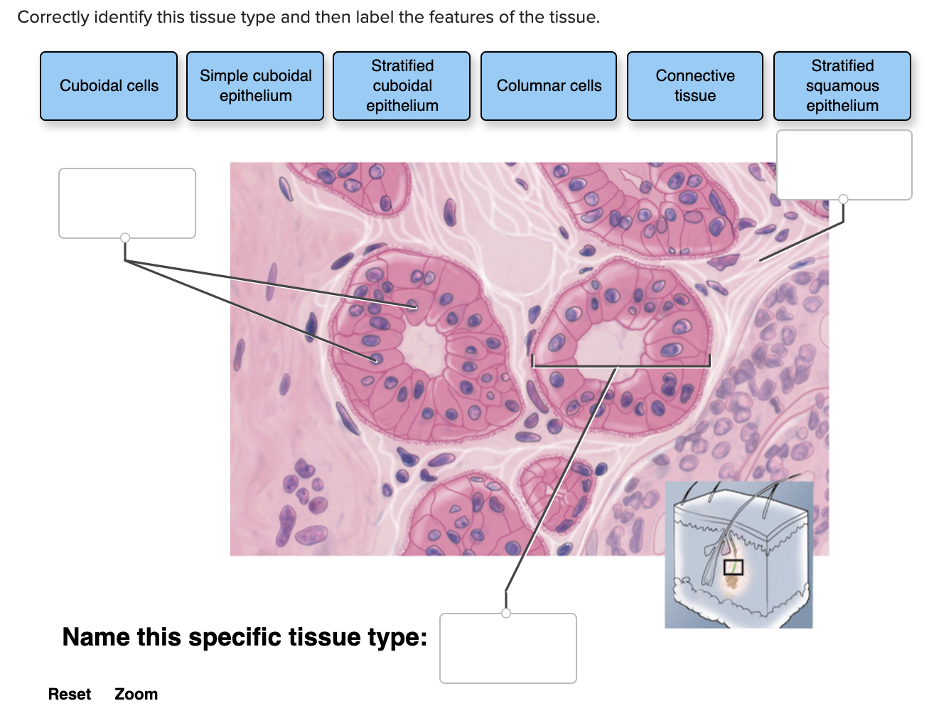
Simple Cuboidal Epithelium Function & Location | What Is ... Simple Cuboidal Epithelium: Labeled Diagram Simple cuboidal epithelial cells are shaped like cubes, and the nucleus of each cell is large and located close to the center of the cell. PDF Tissue Images - PC\|MAC (a) Diagram: Simple squamous (b) Diagram: Simple cuboidal (c) Diagram: Simple columnar Photomicrograph: Simple cuboidal epithelium in kidney tubules (250 ×). Photomicrograph: Simple columnar epithelium of the small intestine (575×). Photomicrograph: Simple squamous epithelium forming part of the alveolar (air sac) walls (275×). Simple squmous epithelium, c.s. - Austin Community College ... Simple squamous epithelium, c.s. (400X) Kidney cortex: This image is the area that was enclosed in a rectangle in the previous image. The dark purple spots are the nuclei of cells, and the cytoplasm is stained a dark pink color. The glomerular capsule is marked with an asterisk (*). Simple Squamous Epithelium Diagram - Quizlet Start studying Simple Squamous Epithelium. Learn vocabulary, terms, and more with flashcards, games, and other study tools.
Simple columnar epithelium Images, Stock Photos & Vectors ...
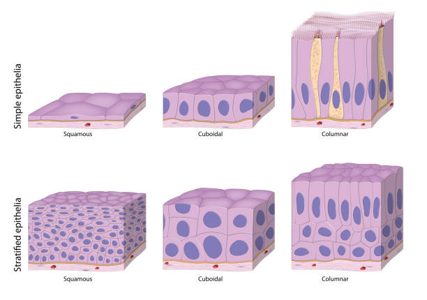
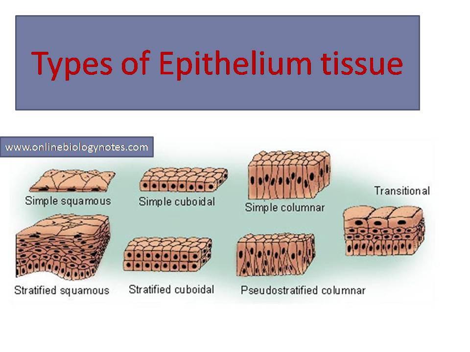
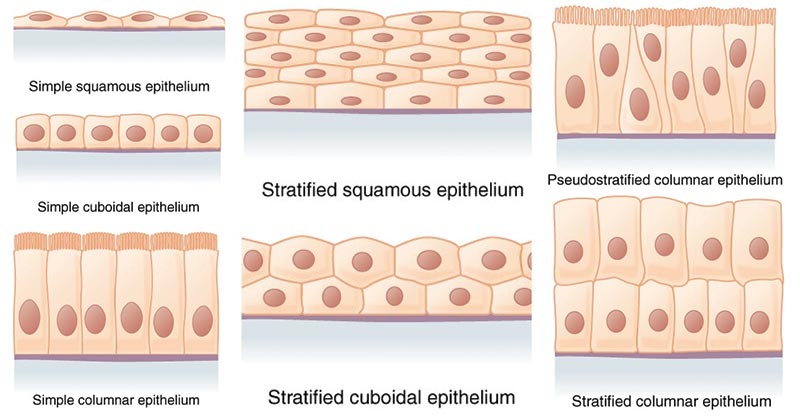
Simple epithelium: Location, function, structure | Kenhub Simple epithelium can be divided into 4 major classes, depending on the shapes of constituent cells. The cells found in this epithelium type are flat and thin, making simple squamous epithelium ideal for lining areas where passive diffusion of gases occur.Areas where it can be found include: skin, capillary walls, glomeruli, pericardial lining, pleural lining, peritoneal cavity lining, and ...

62 Simple Squamous Epithelium Illustrations & Clip Art - iStock
Transitional epithelium- definition, structure, functions ... Structure of the transitional epithelium. Transitional epithelium is an epithelial tissue which in a relaxed state appears as a stratified cuboidal epithelium.; The cells in the transitional epithelium are pear-shaped or round, but as tissue is stretched, cells become flattened, giving the appearance of stratified squamous epithelium.; The cells in the basal layer appear cuboidal or columnar ...
Epithelial Tissue | histology
Duke Histology - Urinary System The parietal layer of Bowman's capsule is also a simple squamous epithelium which transitions to cuboidal epithelium of the proximal convoluted tubule at the urinary pole #210 . Look around under low power to find glomeruli sectioned through the vascular pole. Near the vascular pole will be the distal tubule of the same nephron.

4.2 Epithelial Tissue – Anatomy & Physiology
Label the diagram of simple squamous epithelium Diagram ... Start studying Label the diagram of simple squamous epithelium. Learn vocabulary, terms, and more with flashcards, games, and other study tools.

Introduction and Epithelial Tissues - ppt video online download
LiveInternet @ Статистика и дневники ... Haluaisimme näyttää tässä kuvauksen, mutta avaamasi sivusto ei anna tehdä niin.

Describe the structure and function of different types of ...
Integumentary System- definition, organs, functions, diseases Integumentary system definition, Organs, Structure, Physiology and Functions, Integumentary system diseases. Integumentary system diagram.
Describe various types of epithelial tissues with the class ...
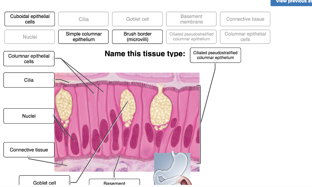
Epithelial Tissue | histology A. Simple columnar epithelium. Slide 29 (small intestine) View Virtual Slide Slide 176 40x (colon, H&E) View Virtual Slide Remember that epithelia line or cover surfaces. In slide 29 and slide 176, this type of epithelium lines the luminal (mucosal) surface of the small and large intestines, respectively. Refer to the diagram at the end of this chapter for the tissue orientation and consult ...
1,119 Simple Squamous Epithelium Stock Photos, Pictures ...
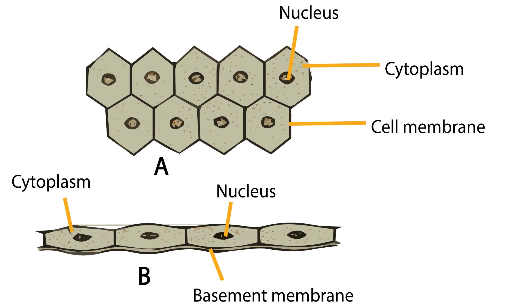
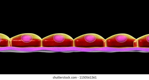
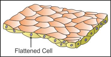
Epithelia: The Histology Guide Squamous. Squamous means scale-like. simple squamous epithelium is a single layer of flat scale-shaped cells. Both the endothelial lining of blood vessels and the mesothelial lining of the body cavities are simple squamous epithelium.. Try to identify the simple squamous epithelia in these pictures.

Study Notes
Simple Columnar Epithelium Labeled Diagram Simple secretory columnar epithelium lines the stomach and uterine schematron.org simple columnar epithelium that lines the intestine also contains a few goblet cells. Simple Columnar Epithelium: A Labeled Diagram and Functions Epithelium is a tissue that lines the internal surface of the body, as well as the internal organs. Simple epithelium is one of the types of epithelium that is divided into simple columnar epithelium, simple squamous epithelium, and simple cuboidal epithelium.
Chapter 4: Tissues, Part 1
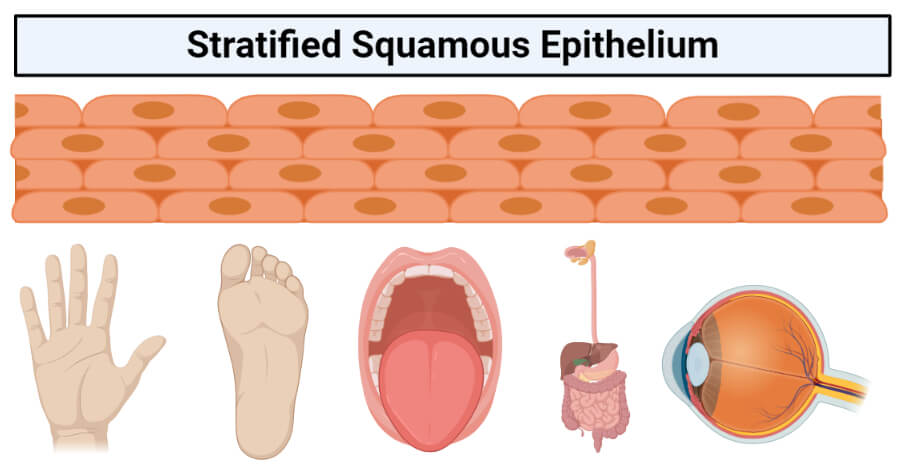
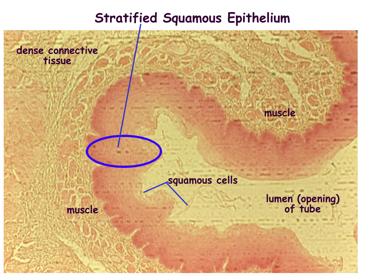
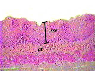
4.2 Epithelial Tissue - Anatomy & Physiology Stratified squamous epithelium is the most common type of stratified epithelium in the human body. The apical cells appear squamous, whereas the basal layer contains either columnar or cuboidal cells. The top layer may be covered with dead cells containing keratin. The skin is an example of a keratinized, stratified squamous epithelium.
Simple Squamous Epithelium
Ruminant Reticulum Histology Slide with Labeled Diagram ... Again, the reticulum microscope labeled image shows the distinguished keratinized stratified squamous epithelium. The propira submucosa of the diagram shows numerous collagen and elastic fibers. Again, the reticulum labeled diagram shows the inner oblique pattern of the smooth muscle layer. The outer smooth muscle layer of the labeled diagram ...

13 Stratified squamous Epithelium ideas | stratified squamous ...
PDF Basic Histo diagrams labelled in colour - 2005 These labelled diagrams should closely follow the current Science courses in histology, anatomy and ... squamous epithelium) ORIGIN: ectoderm lamina propria ... Covers the external surface. blood vessel lined by endothelium (simple squamous epithelium) ORIGIN: mesoderm lumen GENERALISED SECTION epithelium OF THE BODY connective tissue beneath ...
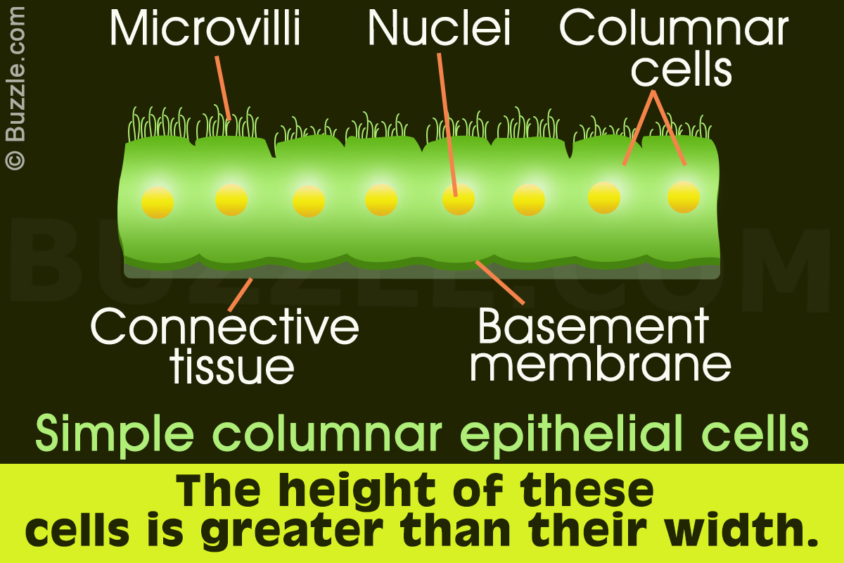
Simple Columnar Epithelium: A Labeled Diagram and Functions ...
Epithelial Tissue: Structure with Diagram, Function, Types ... Simple squamous: Blood vessel lining, air sac lining of lungs: A single layer of flat cells having irregular boundaries: Transport by diffusion and where minimal protection is required: Simple Cuboidal: The tubular lining of kidneys, glandular ducts: A single layer of short cylindrical cells. It may have microvilli as in proximal convoluted tubules

Simple squamous epithelium
Simple Columnar Epithelium Labeled Diagram These labelled diagrams should closely follow the current Science (simple squamous epithelium). ORIGIN: columnar epithelium with goblet cells. TISSUE . Simple Squamous Epithelium Definition Simple squamous epithelium is a single sheet of cells joined together by flat cells cal. Simple columnar epithelium (40X) Primate small intestine.

Pseudostratified Columnar Epithelium Function & Location ...
Types of epithelial tissue: simple, compound and specialized ...

Label the diagram of simple squamous epithelium Diagram | Quizlet

Epithelia: The Histology Guide
Stratified squamous epithelium- structure, functions, examples
Biology 2404: Tissues
Simple squamous epithelium Images, Stock Photos & Vectors ...
Epithelial Tissue - Definition, types, functions, examples

Lab 2 Epithelial tissue | Histology
draw a labelled diagram of squamous epithelial tissue ...

Simple squamous epithelium - Wikipedia

BIOL 237 Class Notes - Histology
Stratified squamous non-keratinized epithelium

Stratified Cuboidal Epithelium Diagram | Quizlet

Stratified squamous epithelium - Wikipedia
Solved Vlew previous atten Simple Simple squamous | Chegg.com

Simple Squamous Epithelium Diagram | Quizlet

3. Types of epithelial cells: simple squamous (top), simple ...

Lab 2 Epithelial tissue | Histology

Simple squamous epithelium - Definition and Examples ...

simple-cuboidal-epithelium | Body tissues, Plasma membrane ...
Solved Correctly identify this tissue type and then label ...

simple squamous epithelium diagram - Google Search | Tissue ...

295 Simple Squamous Epithelium Photos and Premium High Res ...
0 Response to "38 Simple Squamous Epithelium Labeled Diagram"
Post a Comment