41 spinal nerve root diagram
The PNS is a complex system of nerves that branch off from the spinal nerve roots. These nerves travel outside of the spinal canal to the upper extremities ( ... 16/03/2020 · Nerve compression. Compression of the sciatic nerve, or the spinal nerve root, can cause back pain. Pressure from surrounding tissue can irritate the nerve, causing pain, numbness, or tingling ...
Spinal cord within spinal canal Nerve root Intervertebral disc Superior view 10% 9% 8% 23% 18% 23% 9% Motor vehicle accident 23% Motorbike, cycle, pedestrian 23% Low falls 10% 18% Water related 9% Other 8% Struck or collision with person or object 9% Spinal Cord Injury - Traumatic Causes Australia 2008-2009 The Spinal Column Your vertebrae (neck and back …

Spinal nerve root diagram
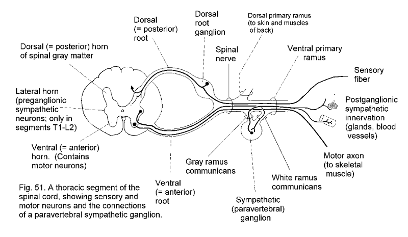
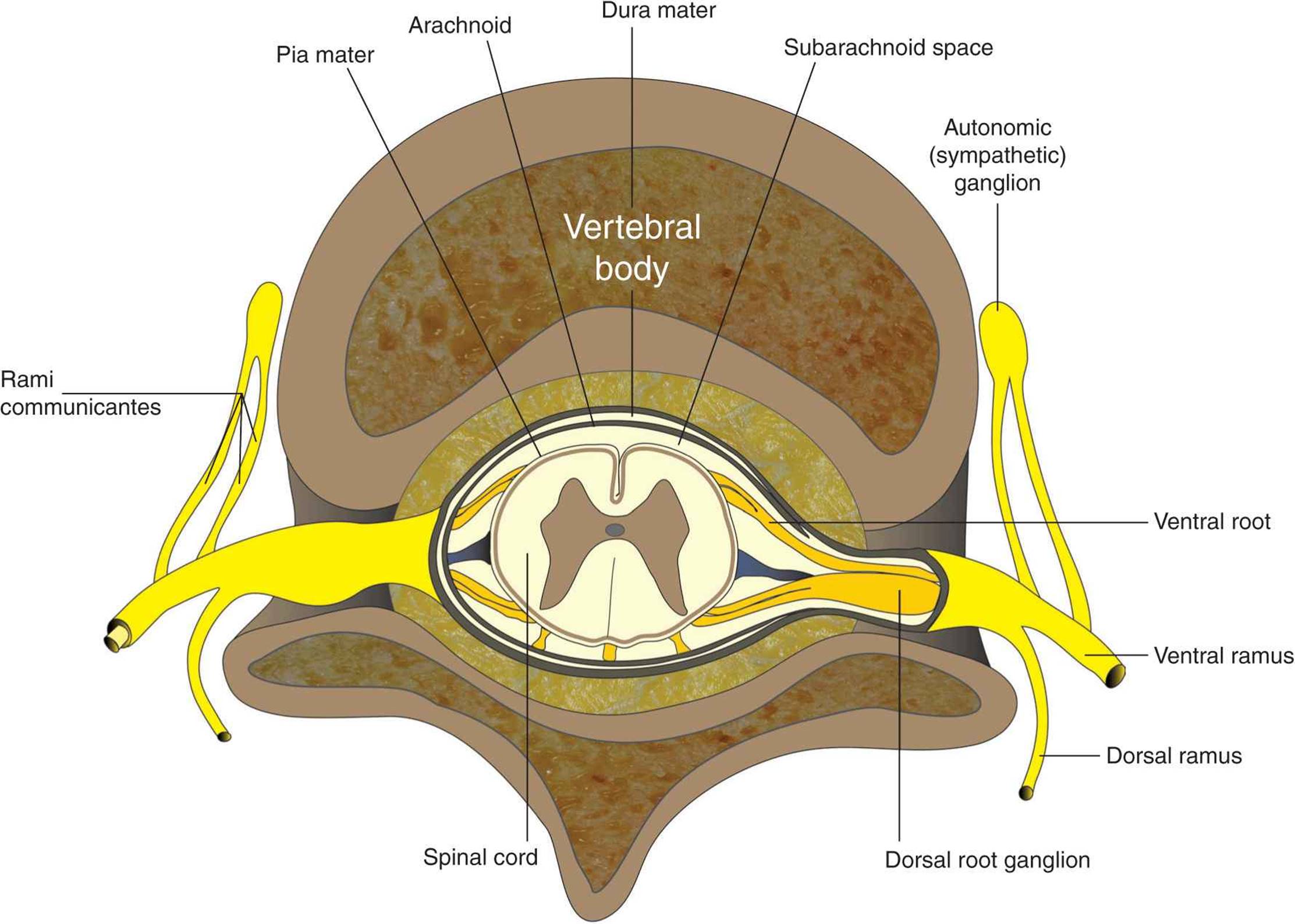
They begin as nerve roots that emerge from a segment of the spinal cord at a specific level. Each spinal cord segment has four roots: an anterior (ventral) ...14 Dec 2018 Nerve Root and Spinal Nerve Anatomy. Each level of the cervical spine has four nerve roots—two on each side—that branch off from the spinal cord. The two types ... The ventral ramus contains nerves that serve the remaining ventral parts of the ... Course and branches of thoracic spinal nerve: This diagram depicts the ...
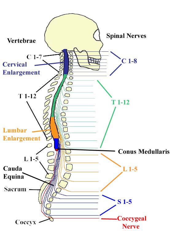
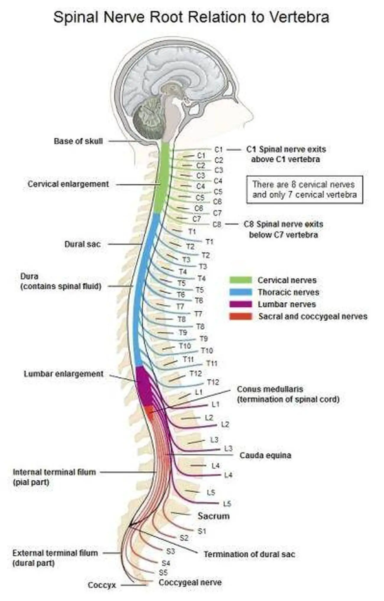
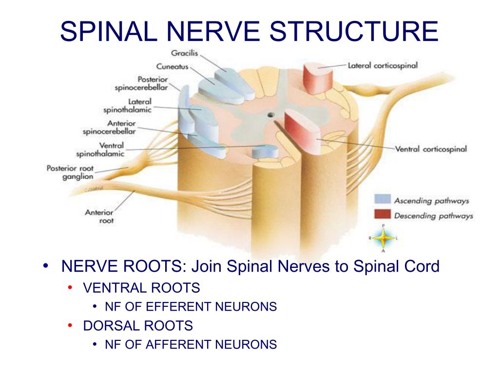
Spinal nerve root diagram. Each spinal nerve is a mixed nerve, formed from the combination of nerve fibers from its dorsal and ventral roots. The dorsal root is the afferent sensory ... Spinal Nerves: In Indian frog, Rana tigrina usually 9 pairs of spinal nerves are found which arise from the spinal cord by two roots, a dorsal or sensory root and a ventral or motor root. Both the dorsal and ventral roots unite immediately after coming out of the neural canal through intervertebral foramen. Dorsal root has a ganglion of nerve ... 14/03/2019 · The sensory root of your trigeminal nerve branches into the ophthalmic, maxillary, and mandibular divisions. The motor root of your trigeminal nerve passes below the … 01/12/2017 · Lateral labeled diagram of the human vertebral spinal column showing vertebrae numbering order and the 5 different regions of the spine. The Atlas is the topmost vertebra, and along with C2, forms the joint connecting the skull and spine. Its chief peculiarity is that it has no body, and this is due to the fact that the body of the atlas has fused with that of the next …
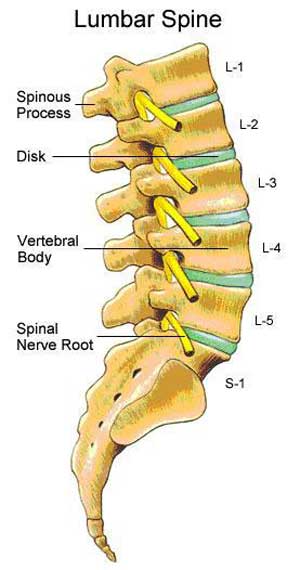
13/06/2019 · Dermatomes are areas of skin that are connected to a single spinal nerve. You have 31 spinal nerves and 30 dermatomes. The exact area that each dermatome covers can be different from person to person. The phrenic nerve is a mixed motor/sensory nerve which originates from the C3-C5 spinal nerves in the neck. The nerve is important for breathing because it provides exclusive motor control of the diaphragm, the primary muscle of respiration. In humans, the right and left phrenic nerves are primarily supplied by the C4 spinal nerve, but there is also contribution from the C3 … Spinal nerve, in vertebrates, any one of many paired peripheral nerves that arise from ... Near the spinal cord each spinal nerve branches into two roots.Related Topics: coccygeal nerve dorsal ramus ...Key People: François MagendieSubjects Of Study: drug action morphine spinal ... 3 Where do the lumbar nerve roots exit, and which root is most likely to be injured in a ... Simplified coronal diagram of lumbosacral plexus, depicted on a ...
Spinal Cord Diagram. Additional Note: The above diagram also showcases the backbone. It contains a total of 24 stacked bones in an adult human. However, at birth, humans have a total of 33 bones in the backbone which eventually fuses to 24 bones. Structure Of Spinal Cord. The Spinal cord runs through a hollow case from the skull enclosed within the vertebral column. … 11/08/2021 · These dorsal root ganglia lie adjacent to the spinal cord and represent the first-order neuron of the spinothalamic tract pathway. The axons of the central process of the first-order neurons enter the spinal cord through the lateral dorsal root entry zone to enter the Lissauer tract and synapses with second-order neurons in the substantia gelatinosa, located in the grey … A dorsal root ganglion (or spinal ganglion; also known as a posterior root ganglion) is a cluster of neurons (a ganglion) in a dorsal root of a spinal nerve.The cell bodies of sensory neurons known as first-order neurons are located in the dorsal root ganglia.. The axons of dorsal root ganglion neurons are known as afferents.In the peripheral nervous system, afferents refer to … The ventral ramus contains nerves that serve the remaining ventral parts of the ... Course and branches of thoracic spinal nerve: This diagram depicts the ...
Image from page 173 of "A healthy body. A textbook on anatomy, physiology, hygiene, alcohol, and narcotics. For use in intermediate grades in public and private schools" (1889)
Nerve Root and Spinal Nerve Anatomy. Each level of the cervical spine has four nerve roots—two on each side—that branch off from the spinal cord. The two types ...

12.3D: Somatic Sensory Pathways - Medicine LibreTexts
They begin as nerve roots that emerge from a segment of the spinal cord at a specific level. Each spinal cord segment has four roots: an anterior (ventral) ...14 Dec 2018

Vertebral Subluxation and Nerve Chart | Spine health ...

Image from page 253 of "Pulmonary consumption, pneumonia, and allied diseases of the lungs : their etiology, pathology and treatment, with a chapter on physical diagnosis" (1901)

Spinal Pathology
Nervous system Flashcards | Easy Notecards

Image from page 552 of "Anatomy, descriptive and surgical" (1887)
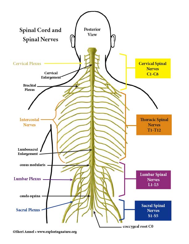
Spinal Cord and Spinal Nerves Diagram
Spinal nerves: Anatomy, roots and function | Kenhub

HumanNervesSpinalFoot - ahuman - Human Spinal Lumbosacral ...

Diagram showing the relationship between spinal nerve ...

The Spinal Cord (Organization of the Central Nervous ...
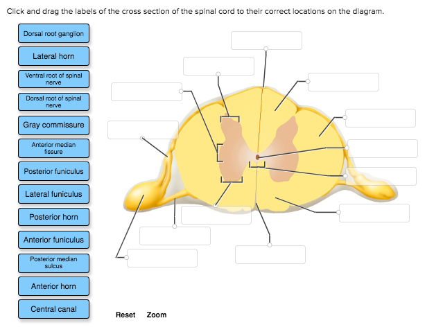
Solved: Click And Drag The Labels Of The Cross Section Of ...

Image from page 95 of "Diseases of the nervous system" (1910)

Image from page 686 of "Elements of human physiology" (1907)
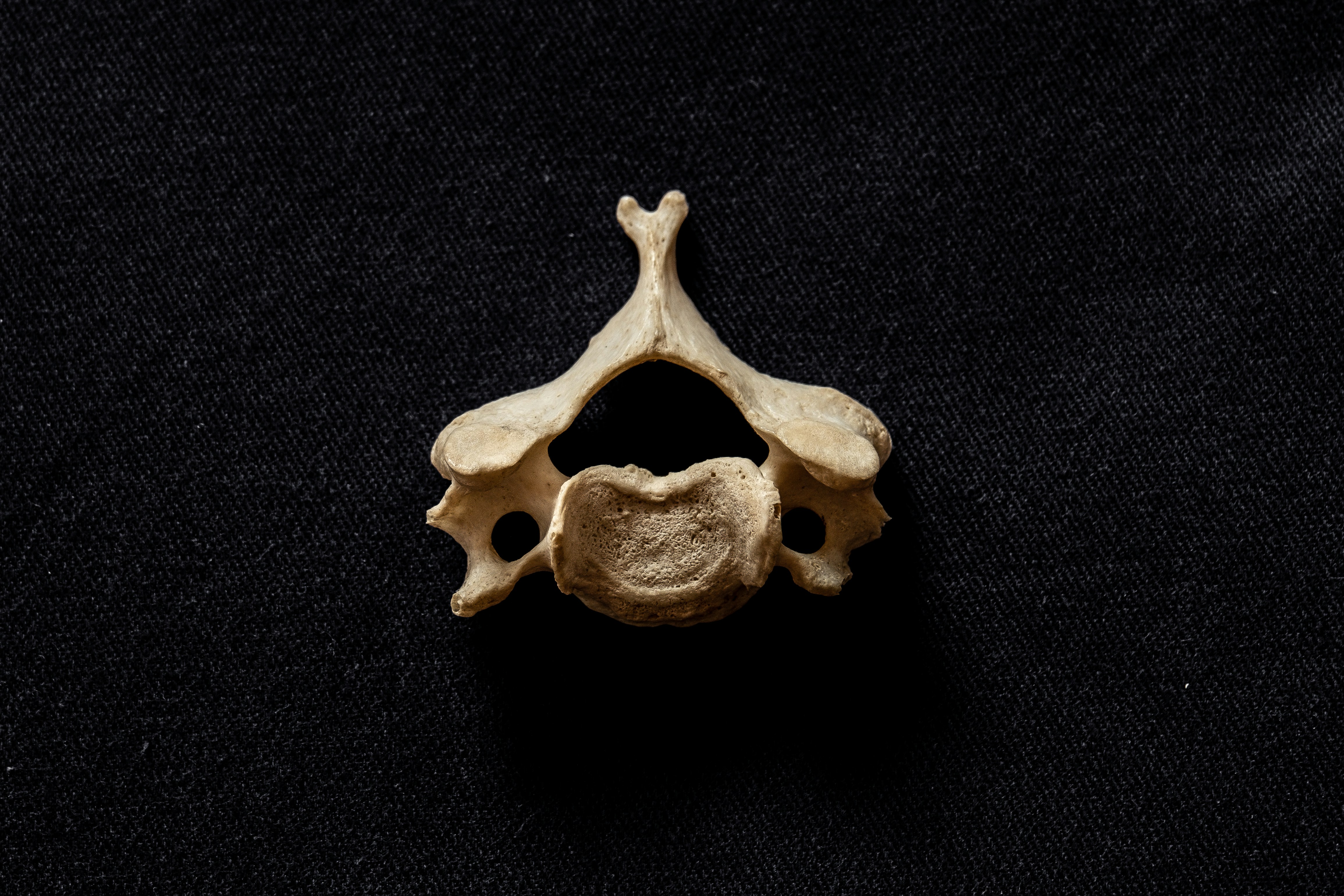
Typical C3-6, human cervical vertebra , bifid spinous process clearly visible

Image from page 611 of "Elements of human physiology" (1907)

A, The dermatomal distribution of the C2-4 nerve root. B ...
Closeup of skeleton pelvic model

Image from page 305 of "The anatomy of the nervous system, from the standpoint of development and function" (1920)
Print Lower Limb Slides & Learning objectives flashcards ...
The structures of a typical spinal nerve Stock Photo - Alamy

Innervation of the lumbar spine. (a) Diagram shows the ...

13.2: The spinal cord is surrounded by three meninges and ...
Peripheral Nervous System: Spinal Nerves and Plexuses
Examining The Spinal Nerve Chart (Neck Pain Support Blog)
Module - Spinal Cord and Spinal Nerve (4 of 14)

Dorsal Root Ganglion | Location, Structure, Histology ...
Intravenous Conscious Sedation for Routine Lumbar Injections

Image from page 50 of "An introductory psychology, with some educational applications" (1911)

MyBiologyPal: Spinal nerves

Print The spinal cord and tracts flashcards | Easy Notecards

Spinal Nerve Chart
Essential Regional Anesthesia Anatomy - Hadzic's ...

Medical Encyclopedia - Structure and Function: Brain ...
Pictures Of Cervical Spinal Nerve

Introducing dye into dorsal root ganglia (DRG) neurons via ...

Image from page 118 of "Nervous and mental diseases" (1911)

Spinal cord injury | The Why Files

November 21st, 2017
PPT - Chapter 14: Peripheral Nervous System PowerPoint ...
0 Response to "41 spinal nerve root diagram"
Post a Comment