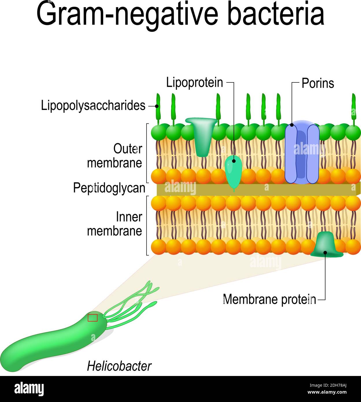
37 gram negative cell wall diagram
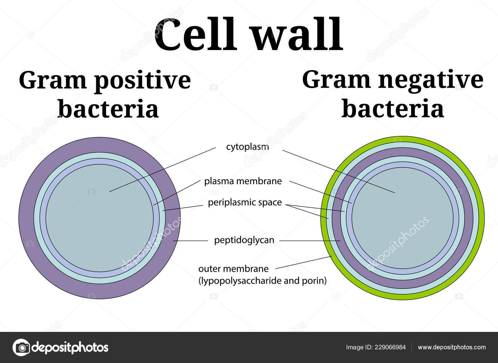
4 Bacteria: Cell Walls . It is important to note that not all bacteria have a cell wall.Having said that though, it is also important to note that most bacteria (about 90%) have a cell wall and they typically have one of two types: a gram positive cell wall or a gram negative cell wall.. The two different cell wall types can be identified in the lab by a differential stain known as the Gram stain.
Gram-negative cell walls are strong enough to withstand ∼3 atm of turgor pressure (), tough enough to endure extreme temperatures and pHs (e.g., Thiobacillus ferrooxidans grows at a pH of ≈1.5) and elastic enough to be capable of expanding several times their normal surface area ().Strong, tough, and elastic … the gram-negative cell wall is a remarkable structure which protects the ...
One such useful classification – if a bacterium is Gram positive or Gram negative - is based on the structure of bacterial cell walls.21 Aug 2019 · Uploaded by Beverly Biology
Gram negative cell wall diagram
Gram staining Procedure. Retains the crystal violet dye and appear purple in colour. Does not retain the crystal violet dye and appear pink in colour. These differences between the Cell Wall of Gram-positive and Gram-negative Bacteria are classified based on their structure, composition of the cell and by the procedure of Gram staining technique.
Download scientific diagram | Diagram demonstrating of the cell wall structure of (a) Grampositive bacterium, (b) Gram-negative bacterium, and (c) mycobacterium. from publication: Differentiation ...
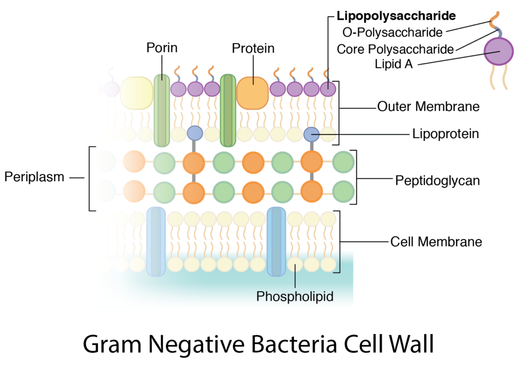
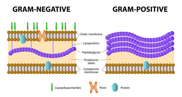
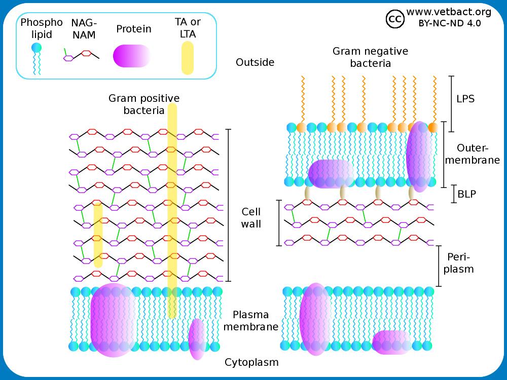
In the Gram-negative Bacteria the cell wall is composed of a single layer of peptidoglycan surrounded by a membranous structure called the outer membrane. The ...
Gram negative cell wall diagram.
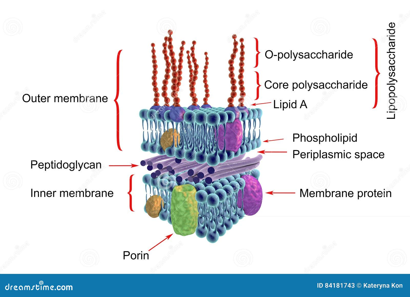
Bacterial Cell Envelope. The Gram Negative Cell Envelope. The diagram above shows a bacterial envelope. It contains an inner membrane, resembling the 'skin' or membrane of an animal cell, in that it is a typical phospholipid bilayer, but then wrapped around this is a cell wall layer of tough but flexible peptidoglycan and then wrapped around ...
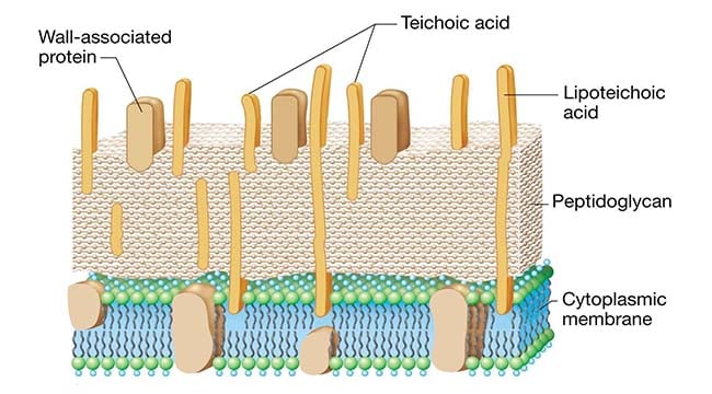
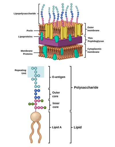
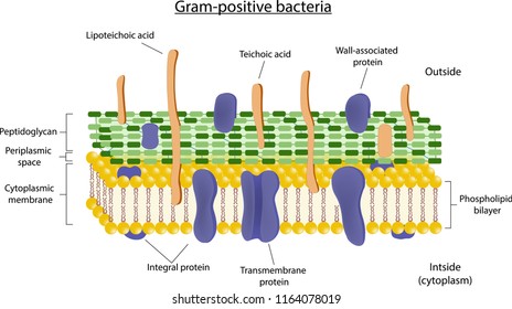
They are characterized by their cell envelopes, which are composed of a thin peptidoglycan cell wall sandwiched between an inner cytoplasmic cell membrane and a ...
Gram-positive Cell Wall. Gram-negative Cell Wall. Outer Membrane. Cytoplasmic Membrane. Membrane Proteins. Porin. RETURN to CELL ENVELOPE . RETURN to CELL DIAGRAM ...
In electron micrographs, the Gram-negative cell wall (Figures 1) appears multilayered. It consists of a thin, inner wall composed of peptidoglycan and an outer membrane. Figure 2.3 B. 1 (left): Electron Micrograph of a Gram-Negative Cell Wall (right) Structure of a Gram-Negative Cell Wall.
The cell wall of gram-negative bacteria is complex having a thin layer of the peptidoglycan layer of 2-7nm and a thick outer membrane of 7-8nm thick. Microscopically, there is a space that is seen between the cell membrane and the cell wall, known as the periplasmic space made up of periplasm.
In addition, they may contain polysaccharide molecules. The gram negative cell wall contains three components that lie outside the peptidoglycan layer: 1. Lipoprotein. 2. Outer membrane and . 3. Lipopolysaccharide. Thus the different results in the gram stain are due to differences in the structure and composition of the cell wall.
Cell wall structures of Gram-positive and Gram-negative bacteria and fungi. (A) Gram-positive bacteria have a single lipid bilayer surrounded by a thick but ...
Download scientific diagram | Schematic structure of Gram-positive and Gram-negative cell walls. Gram-positive cell walls contain only one lipid plasma membrane and a thick peptidoglycan layer ...










:max_bytes(150000):strip_icc()/gram_positive_bacteria-5b7f3032c9e77c0050f88457.jpg)






0 Response to "37 gram negative cell wall diagram"
Post a Comment