41 the diagram below shows a bacterial replication fork and its principal proteins.
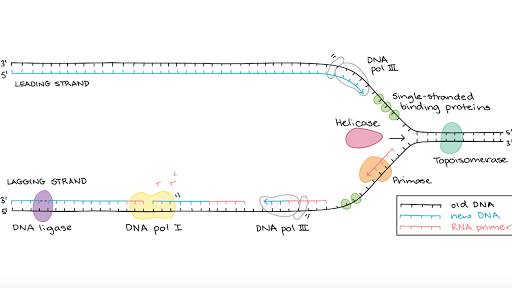
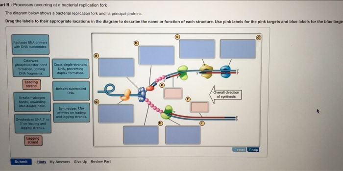
The diagram below shows a bacterial replication fork and its principal proteins. Drag the labels to their appropriate locations in the diagram to describe the name or function of each structure. Use pink labels for the pink targets and blue labels for the blue targets. The diagram below shows a bacterial replication fork and its principal proteins. Drag the labels to their appropriate locations in the diagram to describe the name or function of each structure. Use pink labels for the pink targets and blue labels for the blue targets.
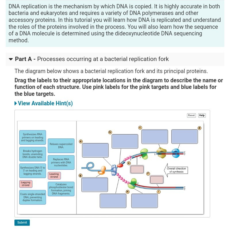
The diagram below shows a bacterial replication fork and its principal proteins. Drag the labels to their appropriate locations in the diagram to describe the name or function of each structure. Use pink labels for the pink targets and blue labels for the blue targets.

The diagram below shows a bacterial replication fork and its principal proteins.
The diagram below shows a bacterial replication fork and its principal proteins. Drag the labels to their appropriate locations in the diagram to describe the name or function of each structure. Use pink labels for the pink targets and blue labels for the blue targets. View Available Hint (s) Reset Help Synthesizes RNA primers on leading and ... The diagram below shows a bacterial replication fork and its principal proteins. Drag the labels to their appropriate locations in the diagram to describe the name or function of each structure. Use pink labels for the pink targets and blue labels for the blue targets The diagram below shows a bacterial replication fork and its principal proteins. Drag the labels to their appropriate locations in the diagram to describe the name or function of each structure. (h)
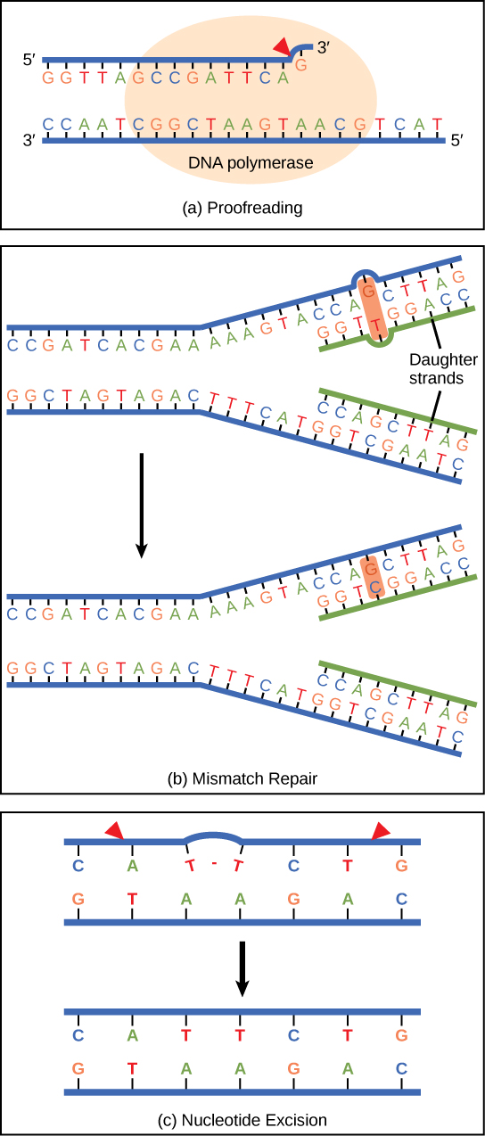
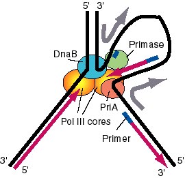
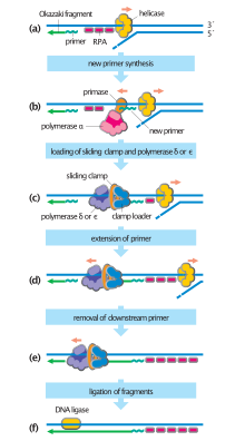
The diagram below shows a bacterial replication fork and its principal proteins.. The diagram below shows a bacterial replication fork and its principal proteins. a. Breaks hydrogen bonds, unwinding DNA double helix Rating: 5 · 1 review The diagram below shows a bacterial replication fork and its principal proteins. Drag the labels to their appropriate locations in the diagram to describe the name or function of each structure. Use pink labels for the pink targets and blue labels for the blue targets. Myosin is a motor protein involved in animal cell cytokinesis. ... The diagram below shows a bacterial replication fork and its principal proteins. The diagram below shows a bacterial replication fork and its principal proteins. The parental dna is shown in dark blue the newly synthesized dna is light blue and the rna primers associated with each strand are red. Use pink labels for the pink targets and blue labels for the blue targets.
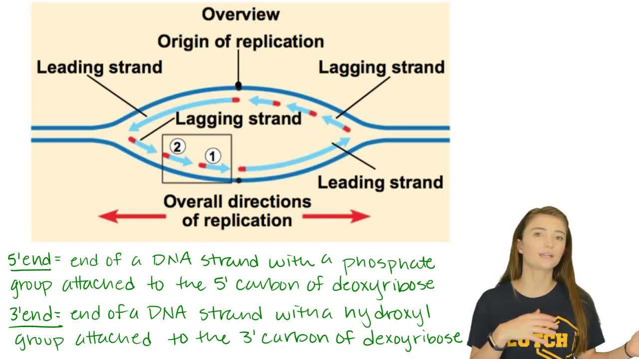
The diagram below shows a replication fork with the two parental DNA strands labeled at their 3' and 5 4/4 (5). The diagram below shows a bacterial replication fork and its principal proteins. Drag the labels to their appropriate locations in the diagram to describe the name or function of each structure. Use pink labels for the pink targets ... The diagram below shows a bacterial replication fork and its principal proteins. Drag the labels to their appropriate locations in the diagram to describe the name or function of each structure. Use pink labels for the pink targets and blue labels for the; Question: The diagram below shows a bacterial replication fork and its principal proteins ... 1 Aug 2018 — Problem: The diagram below shows a bacterial replication fork and its principal proteins. Drag the labels to their appropriate locations in ... The diagram below shows a bacterial replication fork and its principal proteins. Drag the labels to their appropriate locations in the diagram to describe ...
The diagram below shows a bacterial replication fork and its principal proteins. Drag the labels to their appropriate locations in the diagram to describe the name or function of each structure. (h) The diagram below shows a bacterial replication fork and its principal proteins. Drag the labels to their appropriate locations in the diagram to describe the name or function of each structure. Use pink labels for the pink targets and blue labels for the blue targets The diagram below shows a bacterial replication fork and its principal proteins. Drag the labels to their appropriate locations in the diagram to describe the name or function of each structure. Use pink labels for the pink targets and blue labels for the blue targets. View Available Hint (s) Reset Help Synthesizes RNA primers on leading and ...
Dna Replication Microbiology
Journals Uchicago Edu

Origin Of Replication Wikipedia
2
9 2 Dna Replication Concepts Of Biology 1st Canadian Edition

The E Coli Dna Replication Fork Sciencedirect
Chapter 5
Programming For Lovers Chapter 1 Phillip Compeau

Replication Fork Breakage And Restart In Escherichia Coli Microbiology And Molecular Biology Reviews
Machinery Of Dna Replication Springerlink
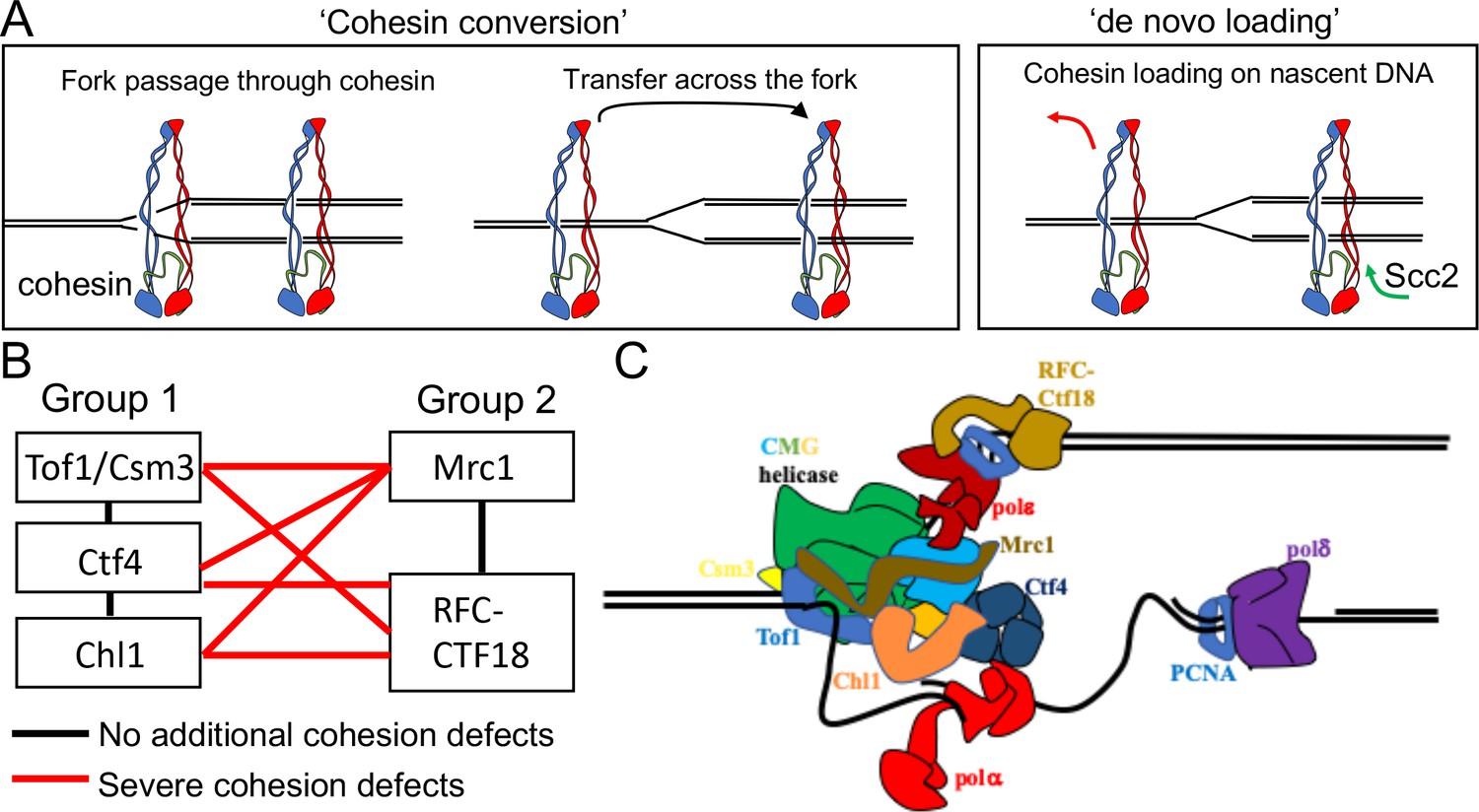
Cohesion Is Established During Dna Replication Utilising Chromosome Associated Cohesin Rings As Well As Those Loaded De Novo Onto Nascent Dnas Elife

Okazaki Fragments An Overview Sciencedirect Topics
Molecular Mechanism Of Dna Replication Article Khan Academy
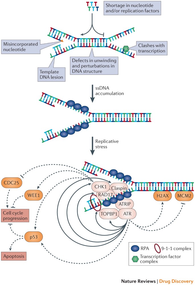
Exploiting Replicative Stress To Treat Cancer Nature Reviews Drug Discovery

Solved The Diagram Below Shows A Bacterial Replication Fork Chegg Com
Chapter 9 Dna Replication Chemistry

Dna Replication Microbiology

Rescuing Replication From Barriers Mechanistic Insights From Single Molecule Studies Molecular And Cellular Biology
Nosborne Com
1

Online Rhs Homework

Arranging Eukaryotic Nuclear Dna Polymerases For Replication Kunkel 2017 Bioessays Wiley Online Library
The Figure Below Is A Dna Replication Bubb Clutch Prep
Solved Processes Occurring At A Bacterial Replication Fork Chegg Com

Asymmetric Histone Inheritance In Asymmetrically Dividing Stem Cells Trends In Genetics
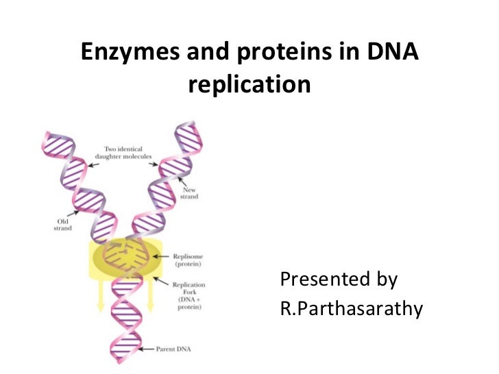
Enzymes And Proteins In Dna Replication
The Diagram Below Shows A Bacterial Replication Fork And Its Principal Proteins Atkinsjewelry

Mind The Replication Gap Royal Society Open Science

Reconstitution Of F Factor Dna Replication In Vitro With Purified Proteins Journal Of Biological Chemistry
Dna Folds Threaten Genetic Stability And Can Be Leveraged For Chemotherapy Rsc Chemical Biology Rsc Publishing Doi 10 1039 D0cb00151a
Drag Each Microdensitometer Graph To
Dna Replication Wikipedia
Ijms Free Full Text R Loops And Its Chro Mates The Strange Case Of Dr Jekyll And Mr Hyde Html

The Dna Damage Response Acts As A Safeguard Against Harmful Dna Rna Hybrids Of Different Origins Embo Reports
2

Stalled Replication Forks Generate A Distinct Mutational Signature In Yeast Pnas
Frontiers Too Much Of A Good Thing How Ectopic Dna Replication Affects Bacterial Replication Dynamics Microbiology
1

The Diagram Below Shows A Bacterial Replic Clutch Prep

Pcr And Molecular Biology Fundamental Principles
Dna Structure Replication Transcription Translation
0 Response to "41 the diagram below shows a bacterial replication fork and its principal proteins."
Post a Comment