39 labeled diagram of neuron
Acetylcholine (ACh) is an organic chemical that functions in the brain and body of many types of animals (including humans) as a neurotransmitter—a chemical message released by nerve cells to send signals to other cells, such as neurons, muscle cells and gland cells. Its name is derived from its chemical structure: it is an ester of acetic acid and choline. 3. How to Draw a Neuron Diagram To learn about the structure of the neurons, the students can use a neuron labeled diagram. The students may follow these steps to make their neuron diagram, but the process is complex: 3.1 How to Draw a Neuron Diagram from Sketch Step 1: First, the students need to draw a circle. Based on it, they need to draw a ...
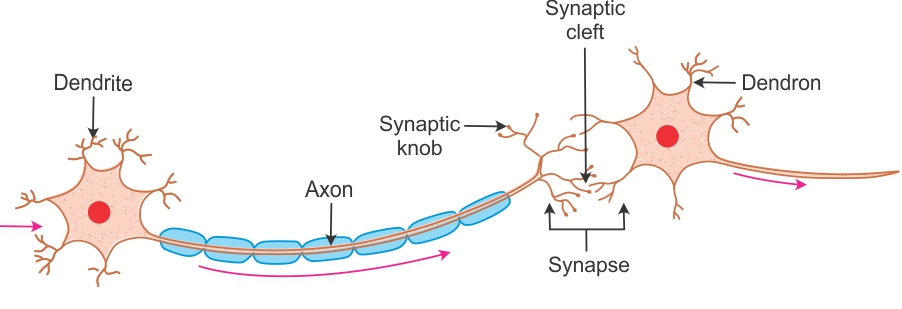
These receive incoming impulses from other neurones. Diagram of a nerve cell A motor neurone. Where two neurones meet there is a small gap called a. Find Motor neuron, detailed and accurate, labeled Stock Vectors and millions of other royalty-free stock photos, illustrations, and vectors in the Shutterstock.
Labeled diagram of neuron
1 answerThe structure of neuron: Nerve cells or neurons are the structural and functional units of the nervous system. It consists of three major parts namely, ... A neuron can be compared to an electrical wire—it transmits a signal from one place to another. Glia can be compared to the workers at the electric company who make sure wires go to the right places, maintain the wires, and take down wires that are broken. Although glia have been compared to workers, recent evidence suggests that also usurp some of the signaling functions of neurons. There ... Click here to get an answer to your question ✍️ Draw neat labelled diagram of neuron.
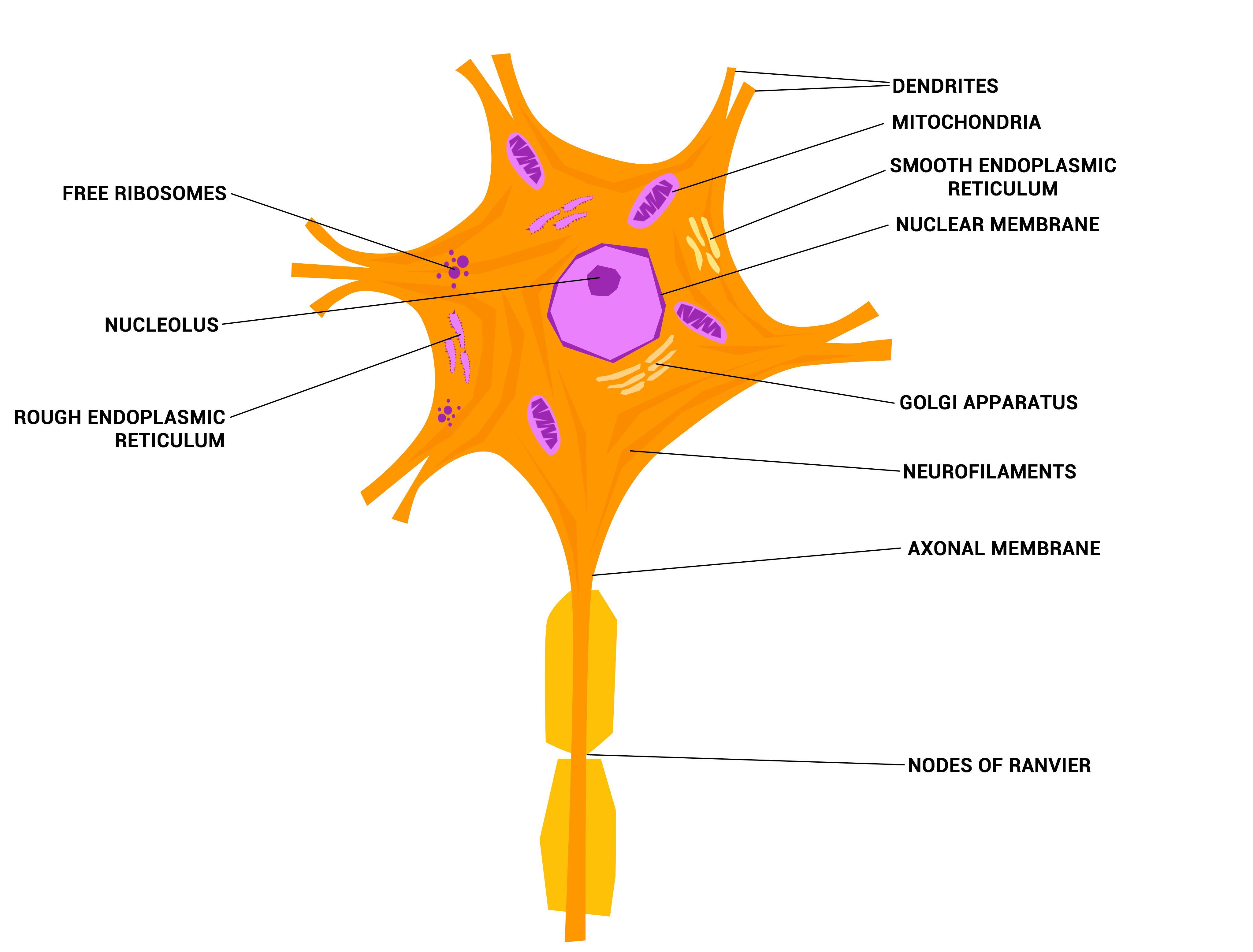
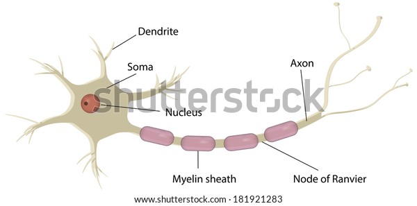
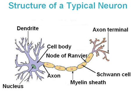
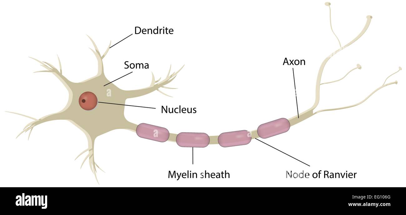
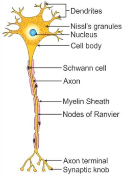
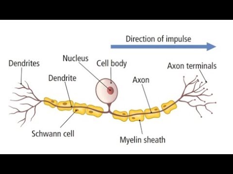
Labeled diagram of neuron. Illustration of Labeled diagram of the neuron, nerve cell that is the main part of the nervous system. vector art, clipart and stock vectors. Hodgkin and Huxley labeled the probability of a gate being in the permissive state as the name of the respective gate. To conform to this method, is exchanged for the name of the gates – m, n, or h. Each channel is defined to have a specific amount of these gates. For example, the sodium channel has three m gates and one h gate [5]. Using ... Draw a labelled diagram of the neuron and describe the structure of the neuron in detail. · 1. Cell body: It forms the cytoplasm of the nerve cell. It is ...1 answer · Top answer: Hint: The cell forms the basic unit of life. Every part of the body has a specialized cell which will perform only a particular activity. They work ... neuron, (1). axon, cell body, dendrites, nucleus, terminal. Unlabeled diagram of a motor neuron (try labeling: axon, dendrite, cell body, myelin, nodes of Ranvier, motor end plate).Read the definitions, then label the neuron diagram below. axon - the long extension of a neuron that carries nerve impulses away from the body of the cell.
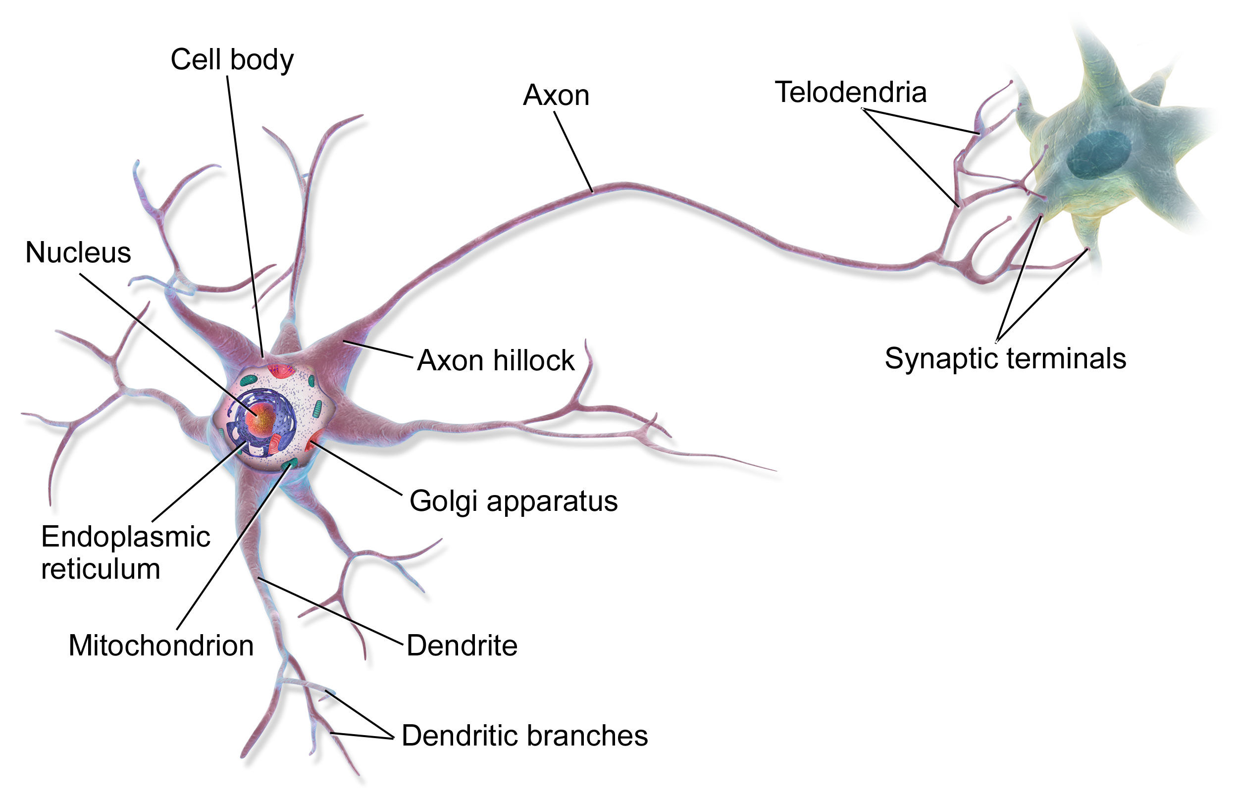
29.09.2021 · (D) Diagram illustrating the coordinated regulation of genes involved in glucose and fatty acid metabolism in HSA-NRTN skeletal muscle compared with WT. See also Figure S4 . Our results show that HSA-NRTN mice recapitulate to a large extent the phenotype observed in skeletal muscle-specific PGC-1ɑ1 transgenic models ( Arnold et al., 2014 ; Gill et al., 2018 ; Handschin et al., 2007 ; … Diagram Of Neuron with Labels · Dendrites–A branch-like structure that functions by receiving messages from other neurons and allow the transmission of messages ...Cell Membrane Definition: Opening and Closing ...Placenta Definition: Introns and ExonsFermentation Definition: Meiosis DefinitionMitochondria Meaning: Rain Water Harvesting ... 15.08.2013 · Each neuron in an MLP takes the weighted sum of its input values. That is, each input value is multiplied by a coefficient, and the results are all summed together. A single MLP neuron is a simple linear classifier, but complex non-linear classifiers can be built by combining these neurons into a network. To me, the RBFN approach is more intuitive than the MLP. An RBFN performs classification 1 answerThe image represents the structure of the neuron and the parts are labelled. expand. Was this answer helpful? upvote 0. downvote 0 ...
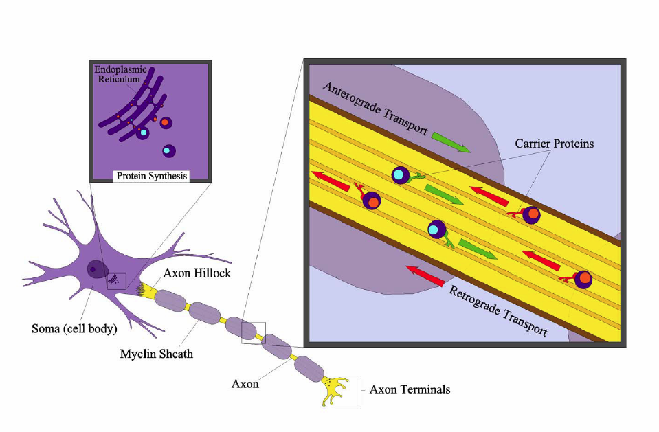
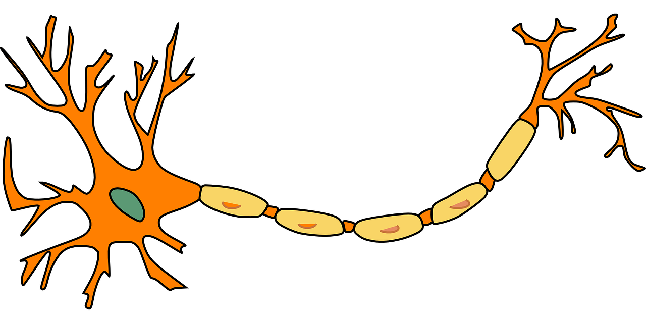
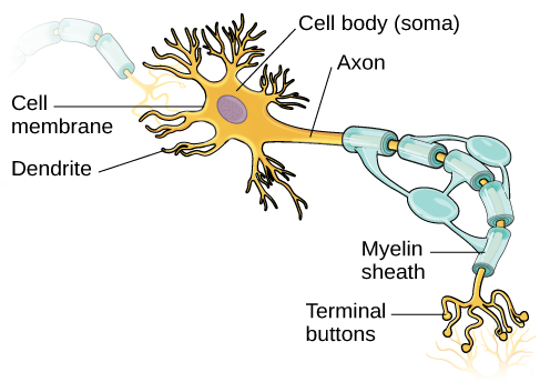
100% (2 ratings) Multipolar motor neurons are found in central nervous system (CNS). Multipolar motor neurons contain multiple dendrite …. View the full answer. Transcribed image text: ures Nucieus Axon FIQURE 25.1 Label this diagram of a mutipolar motor neuron BPR. Draw a labelled diagram of a neuron. Neurons are the fundamental unit of the nervous system.The neuron is a specialized and individual cell, which is also known as the nerve cell. A group of neurons forms a nerve. Dendrites – A branch-like structure that functions by receiving messages from other neurons and allow the transmission of messages ... Click here to get an answer to your question ✍️ Draw a labelled diagram of nerve cell (neuron).1 answer · Top answer: The image represents the labelled image of neuron. Soma: The soma (cell body) is the central mass of the neuron and contains the cell’s nucleus.The nucleus contains the neuron’s DNA and is the site of RNA transcription and translation which produce proteins necessary for the neuron to function properly. The neuron transports proteins from the soma to the axon and dendrites via the activity of microtubule-associated motor proteins.
File Neuron Or Nerve Cell Labelled Image Png Wikimedia Commons
Click here to get an answer to your question ✍️ Draw a labelled diagram of a neuron.
Vektor Stok Nerve Cell Neuron Labeled Diagram Tanpa Royalti 181921283
Parts of a Neuron Diagram. Although they have a characteristic elongated shape, they vary widely in size and properties based on their location and type of functions they perform. While they have the common features of a typical cell, they are structurally and functionally unique from other cells in many ways.
Seer Training Neurons Glial Cells
By contrast, a lower motor neuron lesion to the facial motor nucleus results in paralysis of facial muscles on the same side of the injury. If a cause, such as trauma or infection, cannot be identified (this situation is called idiopathic palsy) this condition is known …

Neuromuscular Junction Vector Illustration Scheme Labeled Cell Infographic Art Print Barewalls Posters Prints Bwc60977910
Neuron and Synapse Labeled Diagram. Human eye anatomy, retina, optic disc artery and vein etc. Structure of a motor neuron. Nerve damage. Human organs thin icons. Synapse and Neuron. Neuron types. Motor neuron. Vector diagram.

Labeled Diagram Of The Neuron Canvas Print Barewalls Posters Prints Bwc31569020
This labelled diagram quiz on Neuron is designed to assess your basic knowledge in Structure and Function of Neuron. Choose the best answer from the four options given. When you've finished answering as many of the questions as you can, scroll down to the bottom of the page and check your answers by clicking ' Score '. Percentage score will be displayed along with right answers.

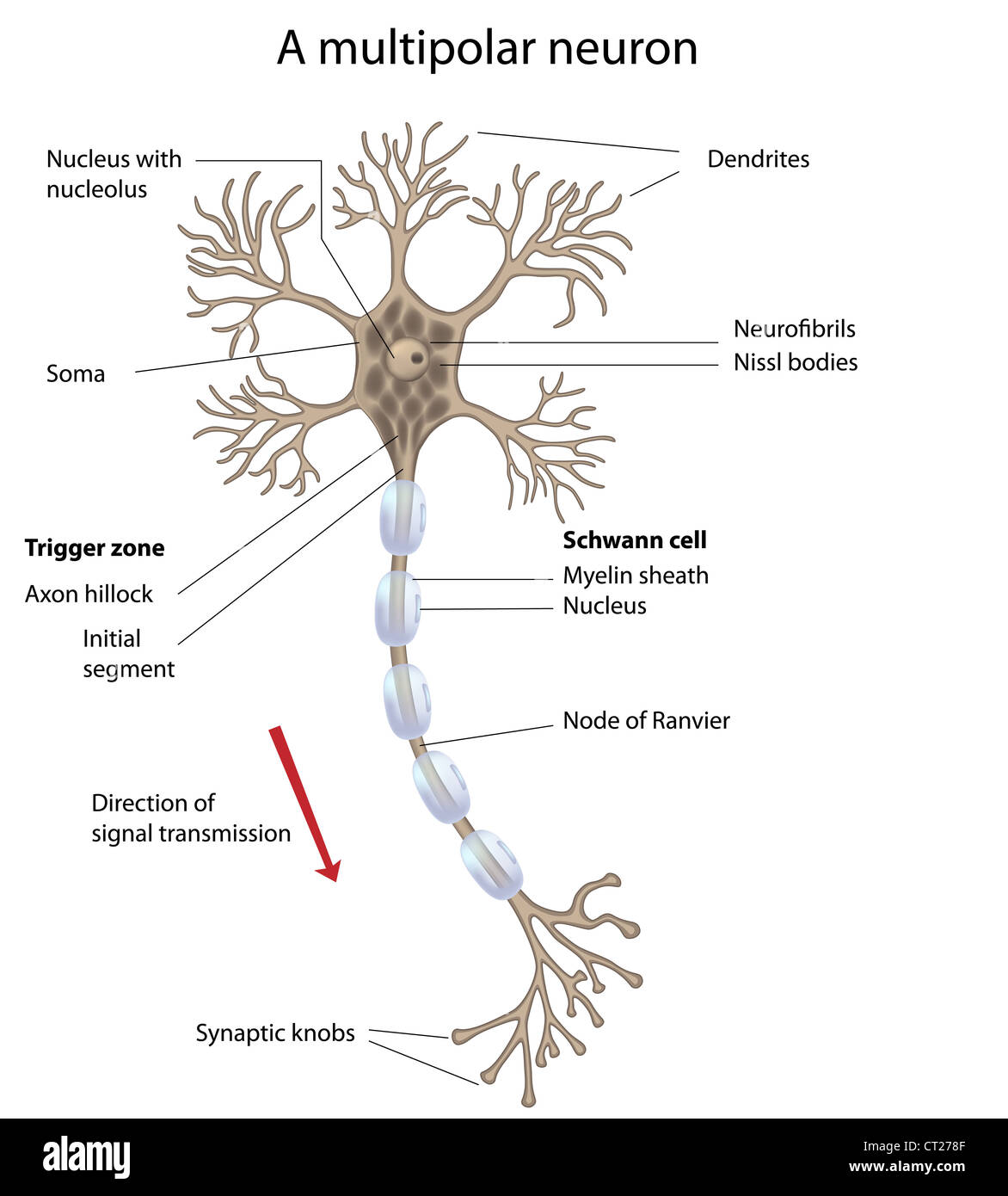
Draw A Labeled Diagram Of A Myelinated Neuron Biology Topperlearning Com A7drhqccc
Click here to get an answer to your question ✍️ Draw neat labelled diagram of neuron.
A Guide To Understand Neuron With Neuron Diagram Edrawmax Online
A neuron can be compared to an electrical wire—it transmits a signal from one place to another. Glia can be compared to the workers at the electric company who make sure wires go to the right places, maintain the wires, and take down wires that are broken. Although glia have been compared to workers, recent evidence suggests that also usurp some of the signaling functions of neurons. There ...
Draw A Labelled Diagram Of Neuron Sarthaks Econnect Largest Online Education Community
1 answerThe structure of neuron: Nerve cells or neurons are the structural and functional units of the nervous system. It consists of three major parts namely, ...

Diagram Of Neuron Anatomy Illustration Of Neuron Anatomy Stock Vector Illustration Of Aorta Biology 203676040

Draw A Labelled Diagram Of A Neuron With A Myelin Sheath

Draw A Labelled Diagram Of A Neuron Scholr
Neuron Labeled Diagram Stock Vector Image Art Alamy

How To Draw Structure Of Neuron Neuron Diagram Labelled Diagram Of Neuron Neuron Cell Youtube Cell Diagram Neuron Diagram Nerve Cell
1

Neuron And Synapse Labeled Diagram Stock Illustration Adobe Stock

Draw A Labelled Diagram Of Neuron Brainly In

Bmsc11001 Label Diagram Neuron Diagram Quizlet

Draw A Labelled Diagram Of A Neuron Tissues Forum
Draw A Labelled Diagram Of A Neuron Cbse Class 11 Learn Cbse Forum
Simple

Label Neuron Anatomy Printout Enchantedlearning Com
Neuron Labeled Diagram Anatomy And Structure
1
Draw A Diagram Of Neuron And Name And Label The Part A Where Information Is Acquired B Through Which Information Travels As An Electric Impulse And C Where The Electric Impulse Must

Diagram Quiz On Neuron Structure And Function Labeling Quiz

Motor Neuron Alila Medical Images
Neuron Structure And Classification
How To Draw Sensory Neuron Well Labelled And Very Easy Youtube
Labeled Neuron Diagram Science Trends

Axon Vector Illustration Labeled Diagram With Explanation And Structure Closeup With Cell Body Terminal Synaptic Cleft Canstock

Draw A Labelled Diagram Of A Neuron Science Shaalaa Com
Neurons Introduction To Psychology
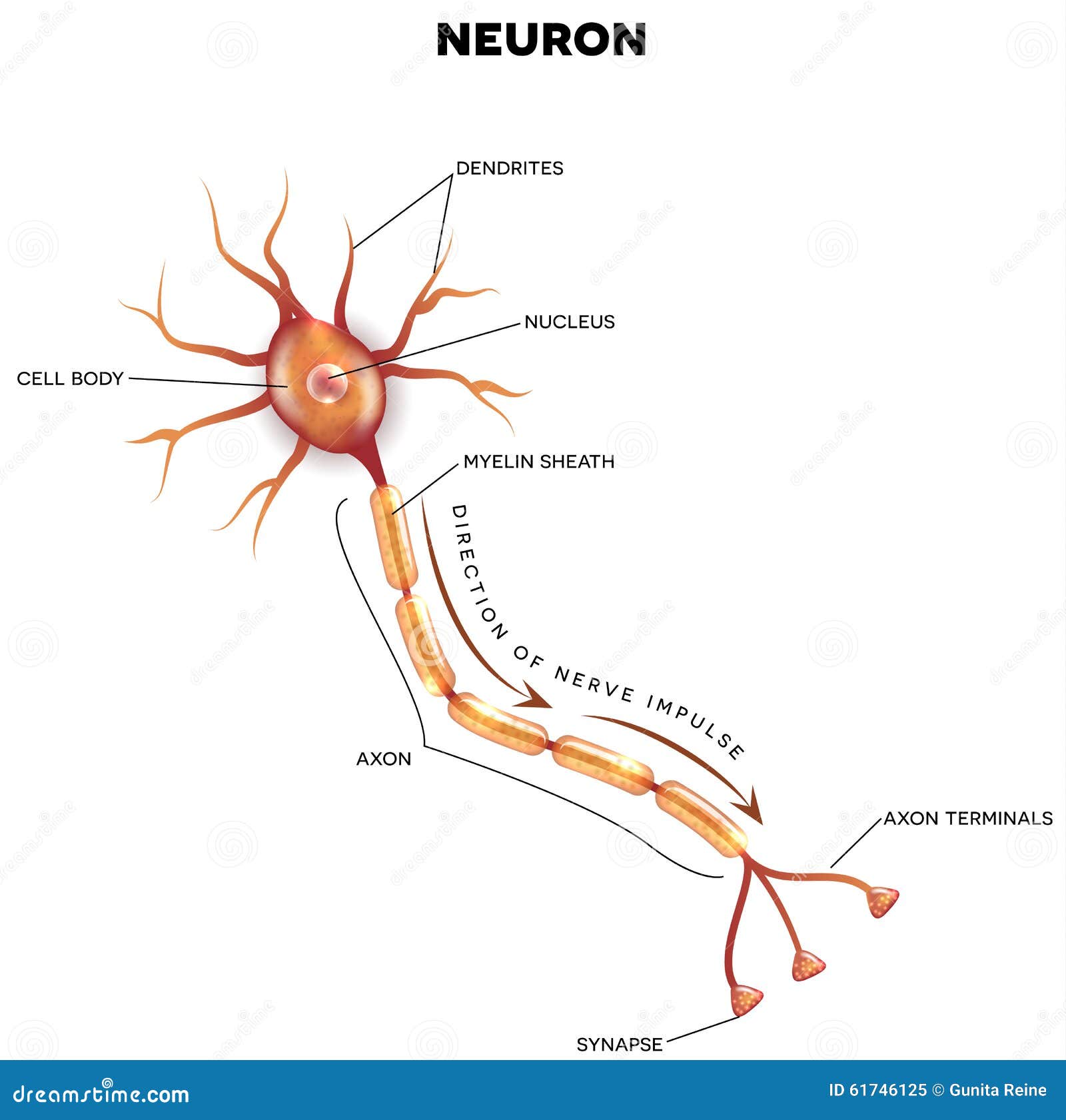
Labeled Diagram Of The Neuron Stock Vector Illustration Of Anatomical Peripheral 61746125

11 Best Neuron Model Project Ideas Neuron Model Neurons Cell Model

Draw A Neat And Labelled Diagram Of The Following A Myelinated Neuron From Biology The Nervous System Class 10 Cbse

Motor Neuron With Myelin And Synapse Alila Medical Images
1
Motor Neuron Labeled Stock Photo Alamy

34 Drawing Of Neuron And Label Of Its Part Labels Database 2020
0 Response to "39 labeled diagram of neuron"
Post a Comment