39 eye to brain connection diagram
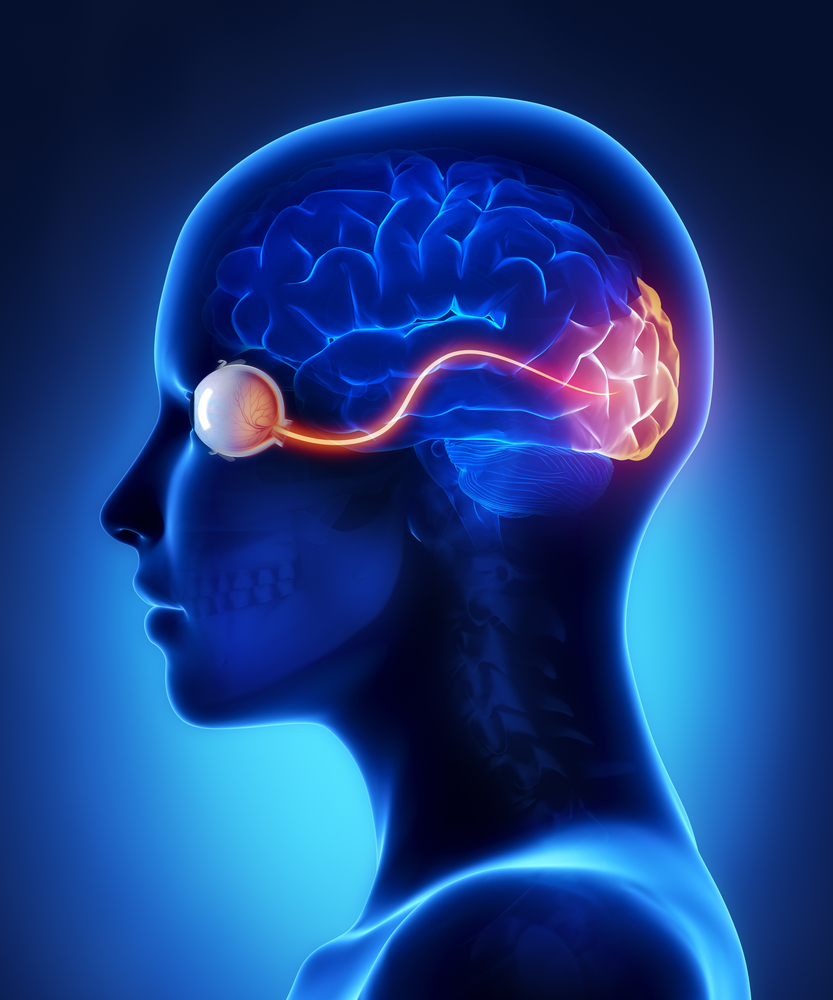
"A wiring diagram of the brain would be a powerful tool to understand diseases of connectivity," said Yuchin Albert Pan, the Commonwealth Research Commercialization Fund Eminent Research Scholar ... Made of nerve cells, the optic nerve is located in the back of the eye. Also known as the second cranial nerve or cranial nerve II, it is the second of several pairs of cranial nerves.It is a bundle of nerve cells that transmits sensory information for vision in the form of electrical impulses from the eye to the brain.
involved in coordinated eye movements, focusing, and papillary responses. Inferior colliculus Part of the midbrain (corpora quadrigemina); contains nerve reflex centers involved in auditory reflexes. Pons Region of brain stem between the midbrain and medulla oblongata; serves as the bridge (connection) between the two regions, and the cerebellum.

Eye to brain connection diagram
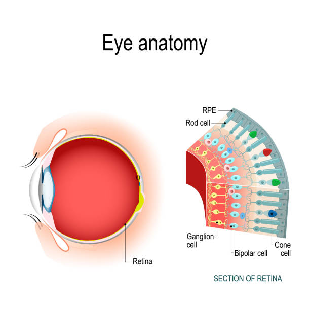
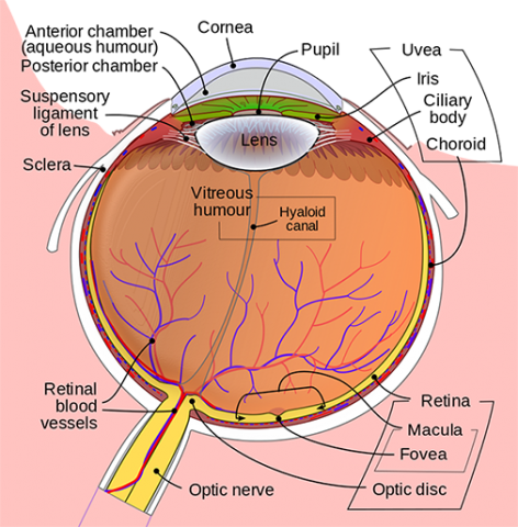
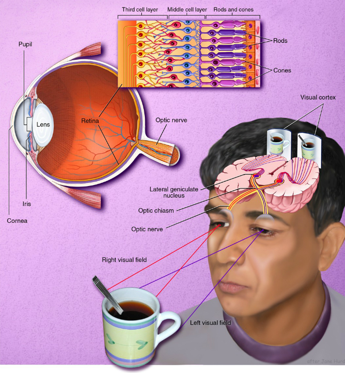
At this point, The light then passes from the lens to the back of the eye which is filled with a clear, gelatinous substance called the vitreous until it reaches the retina, the light-sensitive layer at the back of the eye. The eye-brain connection. Vision is dependent on the connections between the eyes and the brain. A wandering eye can often be corrected surgically, but not until a baby is old enough. In the meantime, one covers an infant's good eye with a patch for a few hours each day so that the brain must rely on signals from the bad eye. This helps the brain develop properly to process signals coming from both eyes. How eye work medical illustration, eye - brain diagram, eye structure and connection with brains. Vector EPS10. Parts of the eye, labeled vector illustration diagram Parts of the eye, labeled vector illustration diagram. Educational beauty and nursing information. Eyelid, eyelashes, pupil, lacrimal gland and other anatomical parts.
Eye to brain connection diagram. In this lesson plan, which is adaptable for grades 4-12, students use BrainPOP resources to explore how the human eye functions. Students will watch a brief movie on the eye and complete a set of graphic organizers identifying the parts of the human eye and their functions. Students will then complete a simple research project on the eye structure abnormalities that cause nearsightedness and ... What is the Third Eye? The pineal gland is a pea-sized gland shaped like a pine cone, located in the vertebrate brain near the hypothalamus and pituitary gland. Also known as the third eye, it is a revered tool of seers and mystics and considered to be the organ of supreme universal connection. The researchers were able to identify most of the 950 neurons included in the new retinal-wiring diagram based on their connections with other neurons, as well as the shape of the neuron. A handful of neurons could not be classified because there was only one of their type, or because only a fragment of the neuron was included in the imaged sample. The Amazing Brain: Toward a Wiring Diagram of Connectivity. ... They have prism glasses that can help with balance, brain exercises, etc as your standard eye doctor doesn't dive deep enough to detect abnormality in eyes that can cause balance/coordination issues from head injury. My child has slowly gained some sense of smell back but not all ...
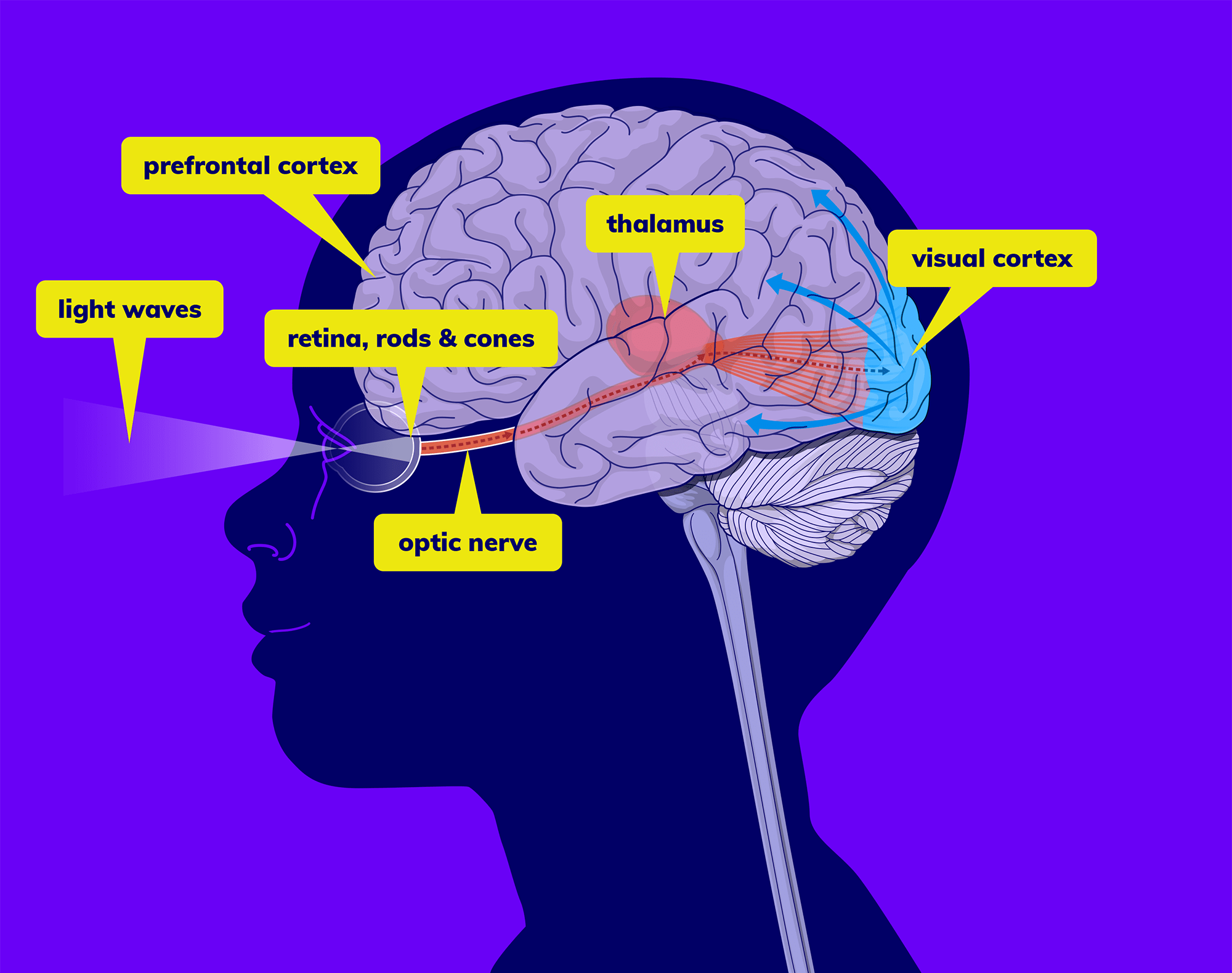
The Eye-Brain Connection. Published 18 Jun 2019; Author Michael W. Richardson Source BrainFacts/SfN Light-sensing cells in your retinas transform light and color into electrical signals. But, to turn that information into a complete picture of the world around you, those signals need to be relayed to multiple areas of the brain quickly and ... Illustration of How eye work medical illustration, eye - brain diagram, eye structure and connection with brains. vector art, clipart and stock vectors. Image 94286109. 52 Ideas For Eye Anatomy Science Eye Anatomy Diagram Of The Eye Human Eye Diagram. Contentseyesvideo Anatomy And Function Of The Eyeearsvideo Ear Anatomy Our Most Important Sensory Receptors Are The Eyes A Ear Anatomy Eye Anatomy Physiology. Eye Diagram Children Eye Diagram Children External Ear Anatomy Ear Anatomy Anatomy. Pin On Useful Things. The cell types in the human brain, for example, showed more diversity. The group also created a wiring diagram of the motor cortex by tracing where the defined cell types project to and from in the brain. Most of the cells formed a complicated network, with multiple connections in different brain areas.
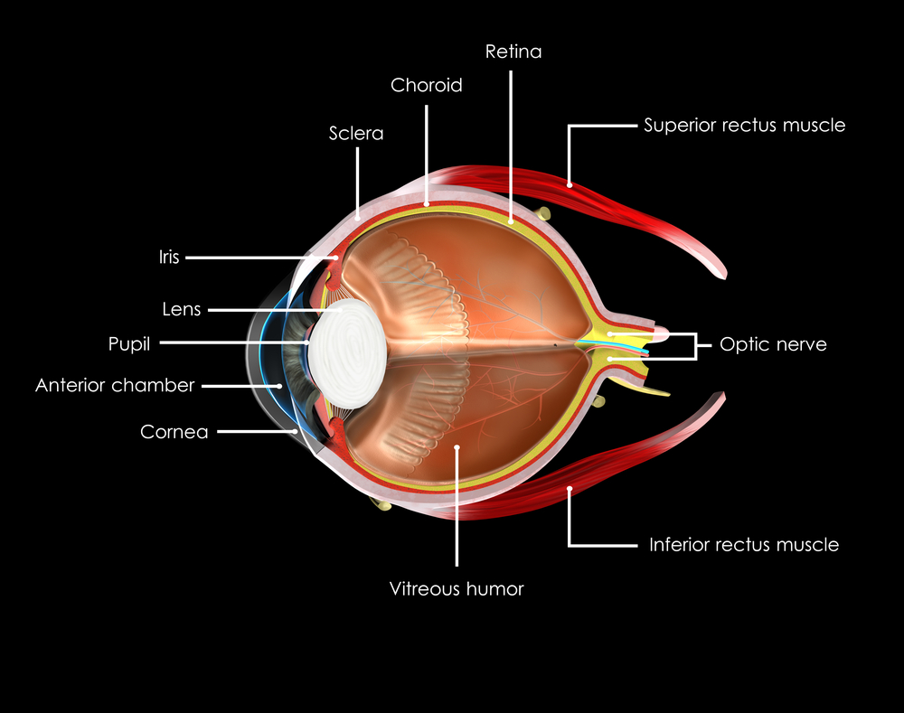
The cornea is located just in front of the iris, which is the colored part of the eye. The main purpose of the cornea is to help focus light as it enters the eye. If one wears contact lenses, the contact lens rests on the cornea. Iris and Pupil. The iris, which is the colored part of the eye, controls the amount of light that enters the eye. The Brain and the Eye. The eye works like a camera. The iris and the pupil control how much light to let into the back of the eye, much like the shutter of a camera. When it is very dark, our pupils get bigger, letting in more light; when it is very bright our irises constrict, letting in very little light. The lens of the eye, like the lens of ... Illustration about How eye work medical illustration, eye - brain diagram, eye structure and connection with brains. Vector EPS10. Illustration of disc, fovea, info - 108512838 Download this How Eye Work Medical Illustration Eye Brain Diagram Eye Structure And Connection With Brains vector illustration now. And search more of iStock's library of royalty-free vector art that features Eye graphics available for quick and easy download.
Membrane
Visualizing the Connections Between Eye and Brain. Featured Neuroscience Visual Neuroscience. · July 2, 2018. Summary: A new study describes the activity of retinal neurons as they deliver visual information to the thalamus, an area of the brain implicated in image processing. Source: BIDMC.

How Eye Work Medical Illustration Eye Brain Diagram Canvas Print Barewalls Posters Prints Bwc54261532
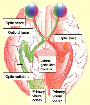
The brain consists of four main segments called lobes. The frontal lobe up front, the parietal lobe on top, the temporal lobe on bottom and the occipital lobe pulling up the rear. All of our senses, thoughts and actions start in one of these lobes. Most visual functions are controlled in the occipital lobe, a small section of the brain near the ...
7 539 Human Eye Anatomy Stock Photos Pictures Royalty Free Images Istock
Citation: Making connections in the eye: Wiring diagram of retinal neurons is first step toward mapping the human brain (2013, August 7) retrieved 9 November 2021 from https://medicalxpress.com ...
The Brain From Top To Bottom
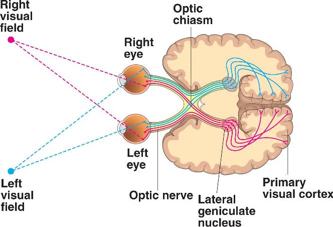
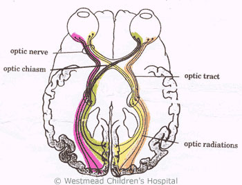
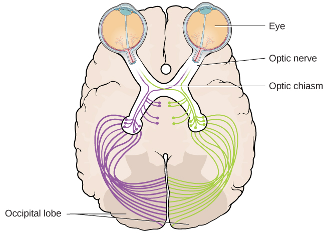
Normally, half the nerves of each eye go to each side of the brain, so that each eye is mapped to both the left hemisphere and the right hemisphere (see wiring diagram), but scans on the German girl showed that retinal nerve fibres that should go to the right hemisphere of the brain diverted to the left.
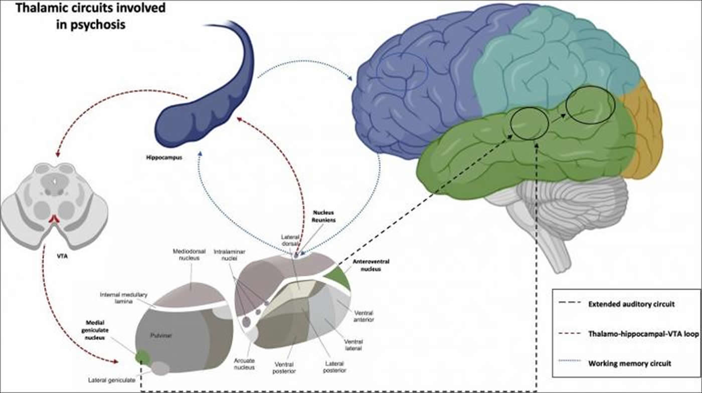
Auditory Hallucinations Rooted In Aberrant Brain Connectivity Neuroscience News
The, clear, gelatinous substance filling the central cavity of the eye. How the Eye Works. The five senses include sight, sound, taste, hearing and touch. Sight, like the other senses is closely related to other parts of our anatomy. The eye is connected to the brain and dependent upon the brain to interpret what we see.

Forebrain Midbrain And Hindbrain Simply Psychology
Human Eye Diagram: Contrary to popular belief, the eyes are not perfectly spherical; instead, it is made up of two separate segments fused together. Explore: Facts About The Eye To understand more in detail about our eye and how our eye functions, we need to look into the structure of the human eye.

Science Of Vision How Do Our Eyes Enable Us To See How It Works
Making connections in the eye: Wiring diagram of retinal neurons is first step toward mapping the human brain Date: August 7, 2013 Source: Massachusetts Institute of Technology

How Eye Work Medical Illustration Eye Brain Diagram Eye Structure And Connection With Brains Stock Illustration Download Image Now Istock
The optic nerve, a cable-like grouping of nerve fibers, connects and transmits visual information from the eye to the brain. The optic nerve is mainly composed of retinal ganglion cell (RGC) axons. In the human eye, the optic nerve receives light signals from about 125 million photoreceptor cells (known as rods and cones) via two intermediate ...
Eye Pictures Anatomy Diagram Body Maps
Optical illusions teach us how our eyes and brain work together to see. You live in a three-dimensional world, so your brain gets clues about depth, shading, lighting, and position to help you interpret what you see. But when you look at a two-dimensional image, your brain can be fooled because it doesn't get the same clues.

How Vision Works Brainhq From Posit Science
The brain is the one who needs to process the visual information, to create order in the world. Eye-brain connection experiment with pilots. A group of pilots was given special glasses that made everyone around them saw upside down. After a few days, their brains corrected the vision, and were able again to see normally, even through those glasses.

Brain And Nervous System For Parents Nemours Kidshealth
PART 1: THE EYE AND THE BRAIN. The display of information must meet the needs of the eye. Someof these needs come from the eye itself, and some come from thebrain. THE EYE, AS WE ALL KNOW, IS A LIGHT-sensitive organ of vision thatpermits us to discriminate among minute variations of shape, color,brightness and distance.
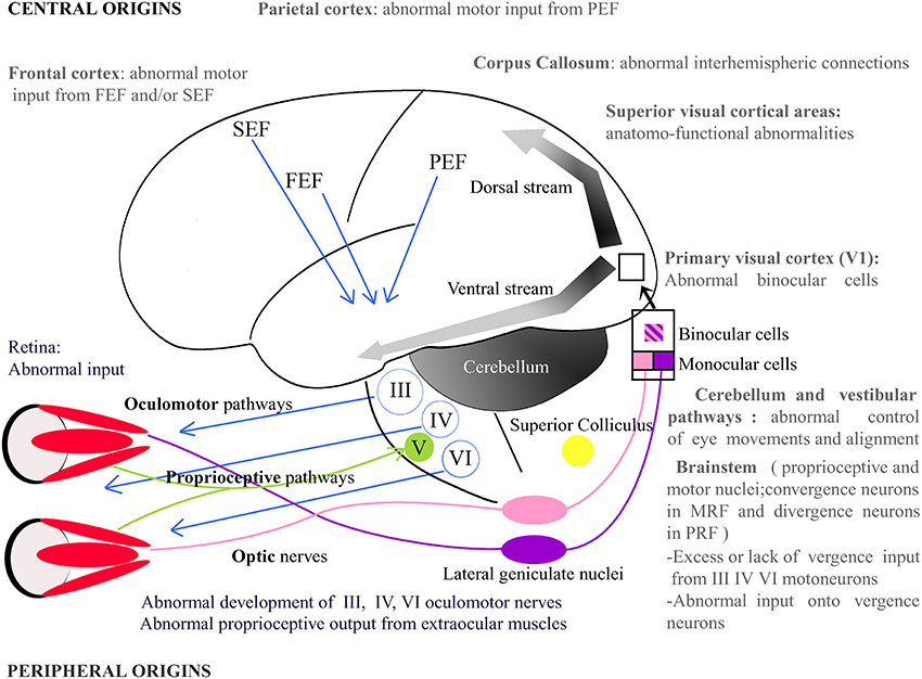
Frontiers Origins Of Strabismus And Loss Of Binocular Vision Frontiers In Integrative Neuroscience
How eye work medical illustration, eye - brain diagram, eye structure and connection with brains. Vector EPS10. Parts of the eye, labeled vector illustration diagram Parts of the eye, labeled vector illustration diagram. Educational beauty and nursing information. Eyelid, eyelashes, pupil, lacrimal gland and other anatomical parts.

How Receptors Of The Eye Conduct Information Via The Optic Nerve Video Lesson Transcript Study Com
A wandering eye can often be corrected surgically, but not until a baby is old enough. In the meantime, one covers an infant's good eye with a patch for a few hours each day so that the brain must rely on signals from the bad eye. This helps the brain develop properly to process signals coming from both eyes.
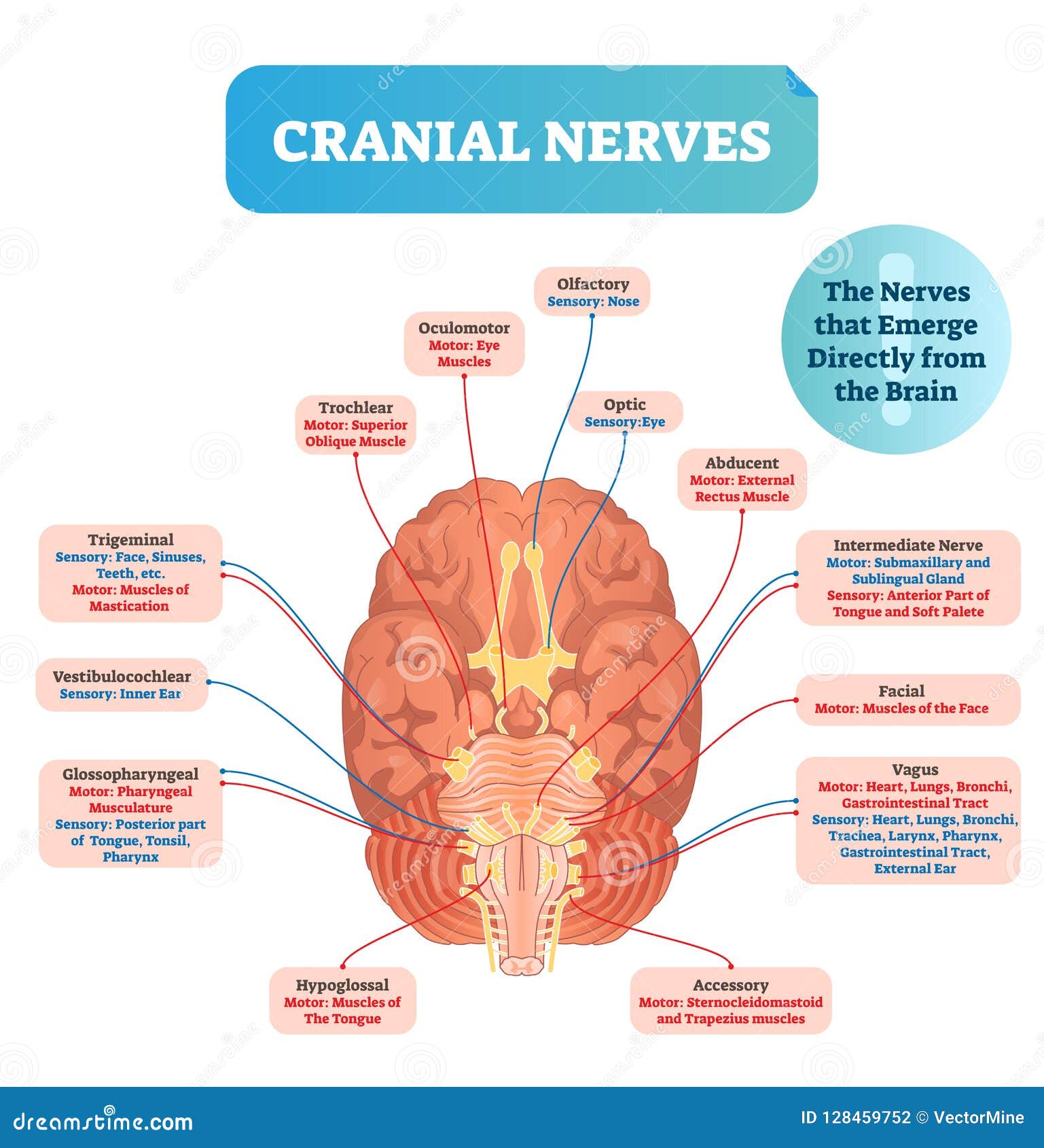
Cranial Nerves Anatomy Names Functions And Mnemonics Kenhub
At this point, The light then passes from the lens to the back of the eye which is filled with a clear, gelatinous substance called the vitreous until it reaches the retina, the light-sensitive layer at the back of the eye. The eye-brain connection. Vision is dependent on the connections between the eyes and the brain.
Kids Health Information Brain Injury Eyes And Vision

Lazy Eye And Amblyopia Precision Family Eye Care
Brain Optic Nerve Stock Illustrations 509 Brain Optic Nerve Stock Illustrations Vectors Clipart Dreamstime
1
/what-are-the-symptoms-of-an-occipital-stroke-3146433-FINAL-814dee4ff85a44e08251a4bcee9536ff.png)
The Effects Of An Occipital Lobe Stroke

What Are The 12 Cranial Nerves And Their Function
Vision Introduction To Psychology

How Do Our Eyes See Color Design Pool

Diagrammatic Representation Of The Interconnection Between Brain Stem Download Scientific Diagram
Anatomy Of The Eye Kellogg Eye Center Michigan Medicine

3 Diagram Of The Human Brain Arrows Indicate The Connection Between Download Scientific Diagram

Perception 3 1 Eye To Brain Youtube
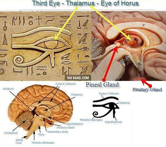
Third Eye The Eye Of Horus Connection To Human Brain 9gag
How We See Color American Museum Of Natural History

How Eye Work Medical Illustration Eye Brain Diagram Eye Structure And Connection With Brains Stock Illustration Download Image Now Istock
The Optic Nerve And Its Visual Link To The Brain Discovery Eye Foundation
Vision It All Starts With Light

Eye And Ear Connection Eyes Problems Eyes Muscle
The Visual Experience Reading 2014
Which Side Of The Brain Does The Optic Nerve Connect To From Each Eye Quora

Basic Structure And Function Of The Nervous System Anatomy And Physiology I
Human Nervous System Diagram How It Works Live Science
Making Connections In The Eye Mit News Massachusetts Institute Of Technology
The Brain And The Eye How They Work Together Discovery Eye Foundation

The Brain And The Eye How They Work Together Golden Eye Optometry
0 Response to "39 eye to brain connection diagram"
Post a Comment