38 pulmonary embolism pathophysiology diagram
17 Aug 2015 — Pulmonary Embolism PART I (Overview) ... Deep Vein Thrombosis - Overview (pathophysiology, treatment, complications). Armando Hasudungan. Diagram of cerebral aneurysm. Cerebral aneurysms are classified both by size and shape. Small aneurysms have a diameter of less than 15 mm. Larger aneurysms include those classified as large (15 to 25 mm), giant (25 to 50 mm), and super-giant (over 50 mm). Berry (saccular) aneurysms. Saccular aneurysms, also known as berry aneurysms, appear as a round outpouching and are the most …
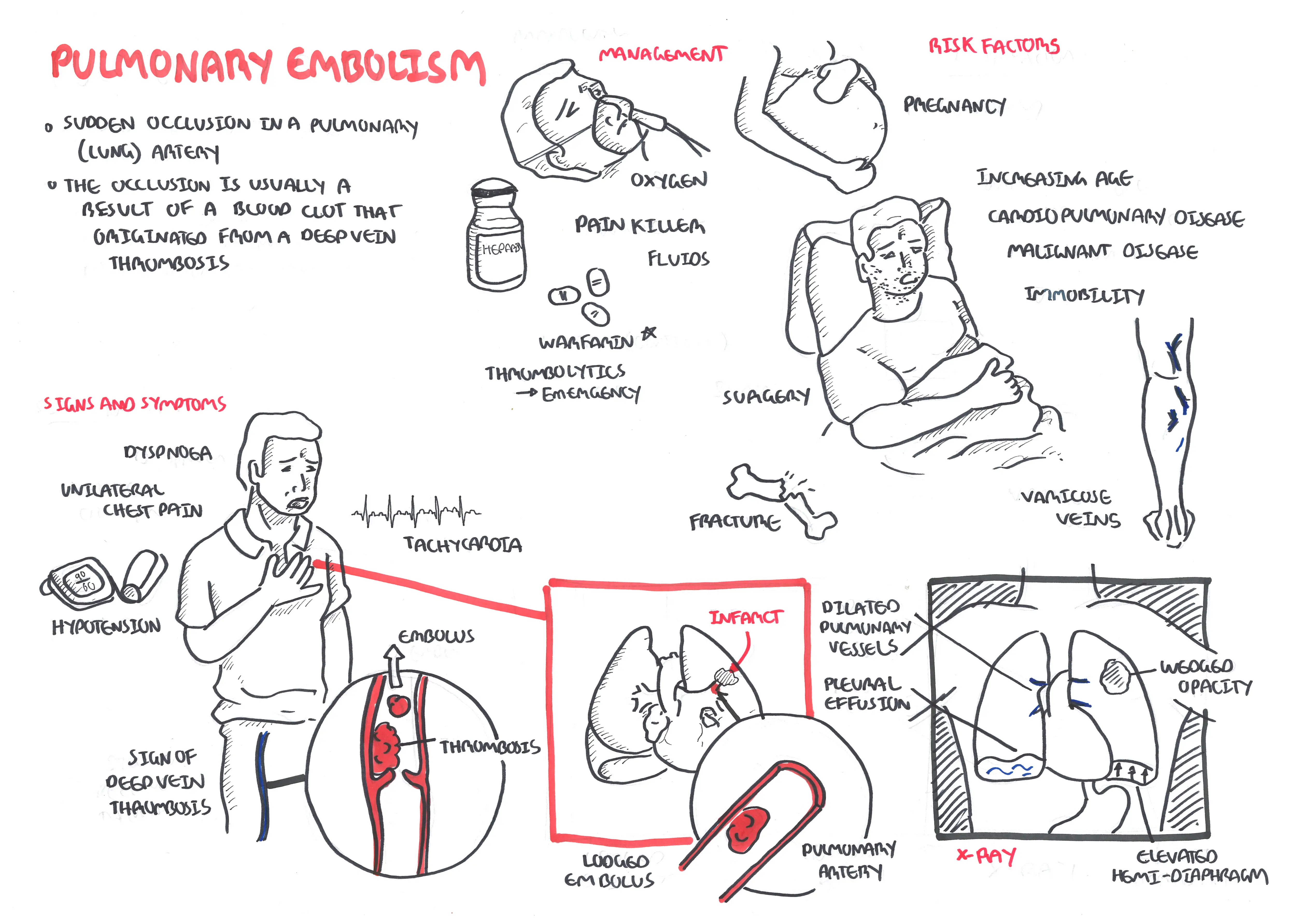
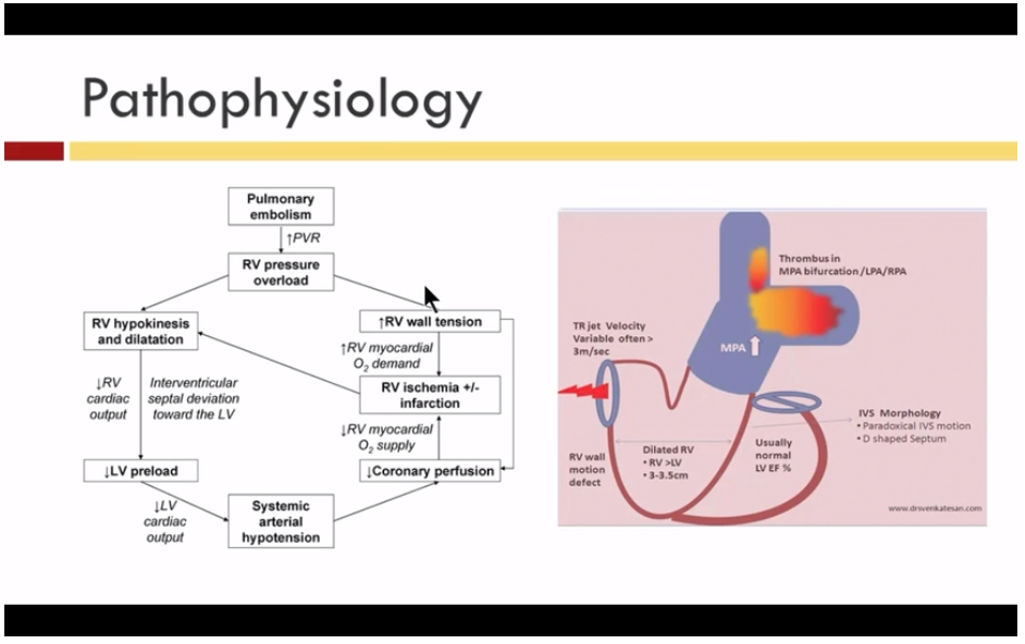
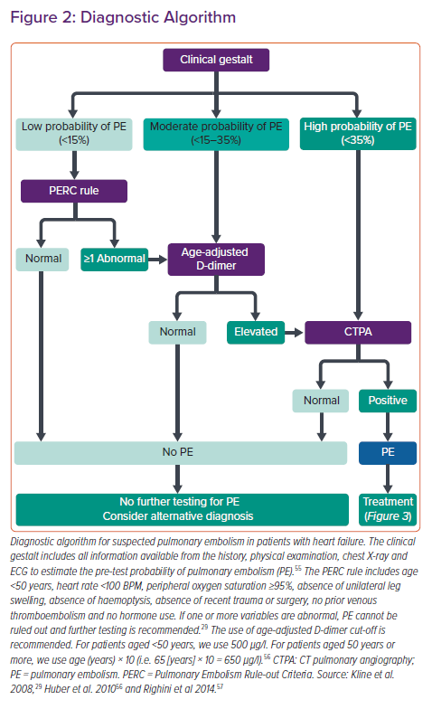
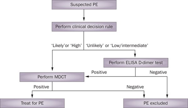
In the first 24 hours, chest x-rays and pulmonary function tests are not definitive for a pulmonary embolism. Oximetry and arterial blood gas typically show hypoxemia. Echocardiography may show right ventricle strain. Serum D-dimer levels will test positive for thrombus degradation by-products; fibrinogen and fibrin.

Pulmonary embolism pathophysiology diagram
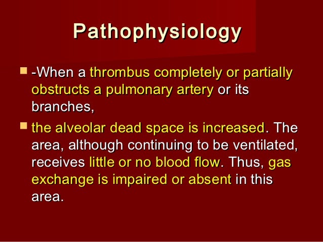
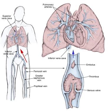
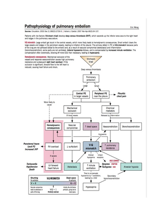
Venous thromboembolism (VTE) » Pathophysiology of pulmonary embolism. Venous thromboembolism (VTE) » Pathophysiology of pulmonary embolism. Posted September 26, 2012 by Eric Wong. Thrombotic pulmonary embolism is not an isolated disease of the chest but a complication of venous thrombosis. Deep venous thrombosis (DVT) and pulmonary embolism are therefore parts of the same process, venous thromboembolism. Evidence of leg DVT is found in about 70% of patients who have sustained a pulmonary embolism; in most of the remainder, it is assumed that the whole thrombus has ... High ventilation/perfusion ratio. High V/Q ratios develop when ventilation is excess in proportion to perfusion. Figure 3 is showing an example of high V/Q ratio in pulmonary embolism (PE). It can produce a dead space like effect.[] Since perfusion is less; removal of CO 2 by high V/Q unit is low. Although the impact of high V/Q unit on blood oxygenation is minimal, it can cause hypoxemia if ...
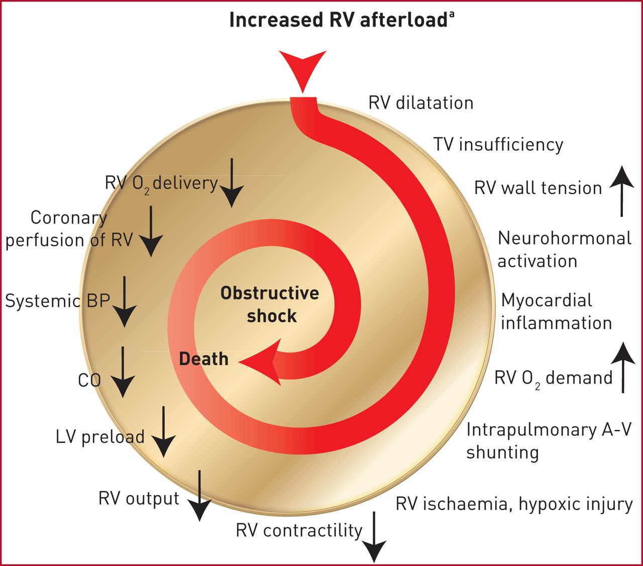
Pulmonary embolism pathophysiology diagram. 18 Sep 2020 — The pathophysiology of pulmonary embolism. Although pulmonary embolism can arise from anywhere in the body, most commonly it arises from the ... Acute pulmonary embolism 1: pathophysiology, clinical presentation, and diagnosis Martin Riedel German Heart Center, Munich, Germany Table 1 Risk factors for venous thromboembolic disease Venous stasis or injury,secondary hypercoagulable states: Immobilisation or other cause of venous stasis—for This New Frontiers article reviews the epidemiology, pathophysiology, diagnosis, treatment, and prevention of pulmonary embolism (PE) in 2 parts. In this first section we summarize the mechanisms of right ventricular dysfunction, arterial hypoxemia, and other abnormalities of gas exchange. For diagnosis, we streamline and expedite the work-up. Pathophysiology of Disease - An Introduction to Clinical Medicine, 7th Ed
Pathophysiology of Pulmonary Embolism. Once deep venous thrombosis develops, clots may dislodge and travel through the venous system and the right side of the ... by DC Sabiston Jr · 1965 · Cited by 65 — Diagrammatic illustration of thrombosis in situ which occurs both proximal and distal to a pulmonary embolus. The proximal thrombosis. Pulmonary embolus is predominantly due to thrombus breaking off from deep veins or from within the right heart, lodging within large or small vessels within the pulmonary vasculature, causing a variable degree of clinical features ranging from asymptomatic through to shock and cardiac arrest. Non-thrombotic causes include air or fat embolism. Outcome is predicated by the degree of right ... emphysema, pulmonary embolism) • To a smaller degree, conditions that alter the membrane’s permeability or increase its thickness (eg, pulmonary fibrosis). The gas used to test the diffusing capacity must be more soluble in bloo d than in lung tis - sue. Ideally, the amount of gas entering the blood should be limited by the lungs’ ability to
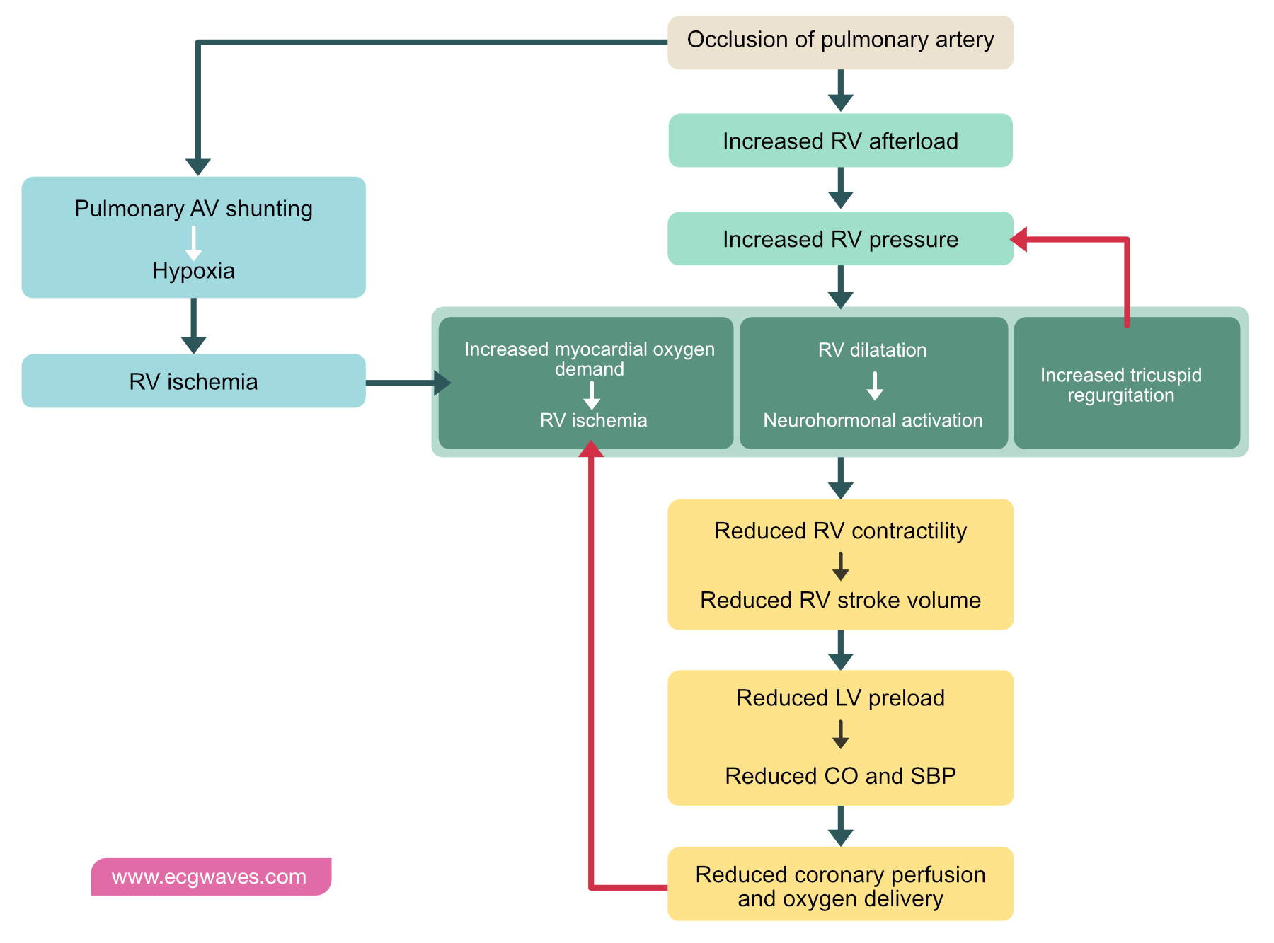
Pulmonary embolism (PE) occurs when there is an acute obstruction of the pulmonary artery or one of its branches. It is commonly caused by a venous thrombus that has dislodged from its site of formation and embolized to the arterial blood supply of one of the lungs. The process of clot formation and embolization is termed thromboembolism. 23 Sep 2019 — Please read and agree to the disclaimer before watching this video.. An embolism is the lodging of an embolus, a blockage-causing piece of ... Pulmonary embolism (PE) is responsible for approximately 100,000 to 200,000 deaths in the United States each year. With a diverse range of clinical presentations from asymptomatic to death, diagnosing PE can be challenging. Various resources are available, ... 28.02.2020 · Pulmonary embolism: Tricuspidal regurgitation: Pneumothorax: Pericardial effusion : Pathophysiological differences between HFpEF and HFrEF. Only in HFrEF but not in HFpEF evidence-based therapy offers improvement in symptoms and prognosis. These differences highlight the need for understanding the differences in the pathophysiology between HFrEF and HFpEF which might also …
Jaypeedigital Ebook Reader
A high incidence of thromboembolic events, including pulmonary embolism (PE), has been observed in COVID-19, which suggests that COVID-19 may induce intravascular coagulopathy [3–6]. PE may be a direct cause of death in patients with COVID-19, despite the use of antithrombotic prophylaxis [4, 6, 7].
Pulmonary Embolism Pe Pulmonary Disorders Msd Manual Professional Edition
High ventilation/perfusion ratio. High V/Q ratios develop when ventilation is excess in proportion to perfusion. Figure 3 is showing an example of high V/Q ratio in pulmonary embolism (PE). It can produce a dead space like effect.[] Since perfusion is less; removal of CO 2 by high V/Q unit is low. Although the impact of high V/Q unit on blood oxygenation is minimal, it can cause hypoxemia if ...
Ojs Uph Edu
Thrombotic pulmonary embolism is not an isolated disease of the chest but a complication of venous thrombosis. Deep venous thrombosis (DVT) and pulmonary embolism are therefore parts of the same process, venous thromboembolism. Evidence of leg DVT is found in about 70% of patients who have sustained a pulmonary embolism; in most of the remainder, it is assumed that the whole thrombus has ...

92 Pulmonary Embolism For The Internist The Curbsiders
Venous thromboembolism (VTE) » Pathophysiology of pulmonary embolism. Venous thromboembolism (VTE) » Pathophysiology of pulmonary embolism. Posted September 26, 2012 by Eric Wong.
Pulmonary Embolism Armando Hasudungan

Pathophysiology Of Pulmonary Embolism Effects Of Grepmed

Survival Benefit Of Obese Patients With Pulmonary Embolism Sciencedirect
Massive Pulmonary Embolism Heart Failure A Review Of Clinical Status And Meta Analyses Of Clinical Scoring System And D Dimer And Thrombolytic And Anticoagulation Therapies
Pathophysiology Of Right Ventricular Failure In Acute Pulmonary Embolism And Chronic Thromboembolic Pulmonary Hypertension A Pictorial Essay For The Interventional Radiologist Insights Into Imaging Full Text
Dr Kon Va Ecmo For Massive Pulmonary Embolism From The Maryland Critical Care Project Tom Wade Md

Pathophysiology Of High Risk Pulmonary Embolism Pe And Intracerebral Download Scientific Diagram

Nonthrombotic Pulmonary Embolism Air Amniotic Fluid Fat Tumor Pulmonology Advisor
Pulmonary Embolism Pe Causes Symptoms Diagnosis Treatment

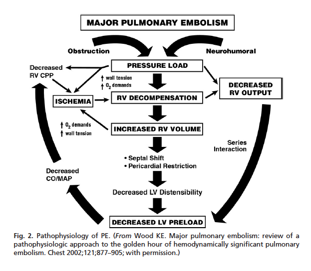
Major Pulmonary Embolism Review Of A Pathophysiologic Approach To The Golden Hour Of Hemodynamically Significant Pulmonary Embolism Sciencedirect

The Pathophysiology Of Chronic Thromboembolic Pulmonary Hypertension European Respiratory Society
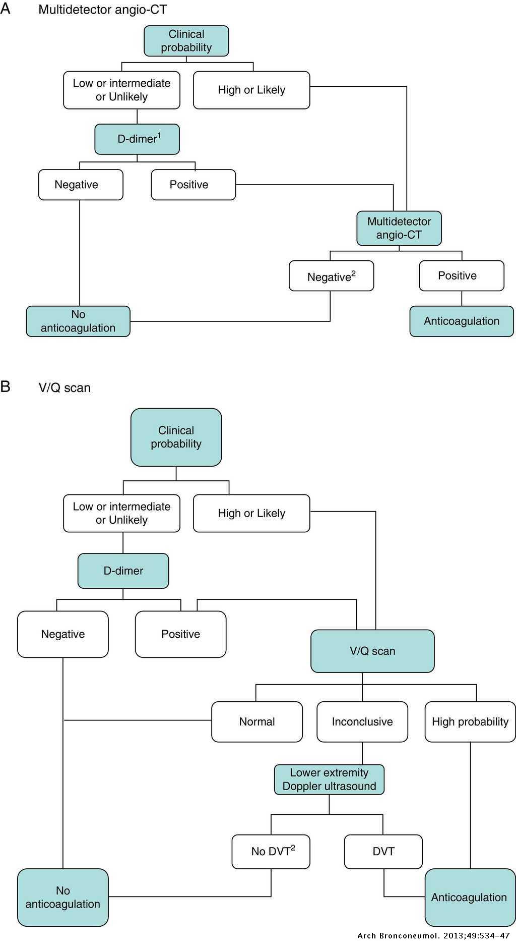
National Consensus On The Diagnosis Risk Stratification And Treatment Of Patients With Pulmonary Embolism Archivos De Bronconeumologia

Massive Pulmonary Embolism Clinical Gate

Pulmonary Embolism Civetta Taylor Kirby S Critical Care 4th Edition

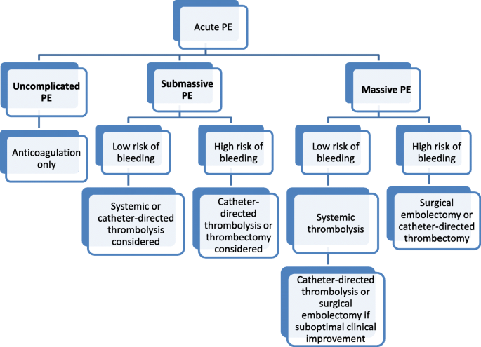
Figure 1 From Management Of Submassive Pulmonary Embolism Semantic Scholar
Pulmonary Embolism

Continuing Professional Development Pulmonary Embolism In Pre Hospital Care Journal Of Paramedic Practice

Pulmonary Embolism
Pulmonary Embolism Heart Failure Cfr Journal
Ocw Ui Ac Id

Flow Diagram Pe Pulmonary Embolism Download Scientific Diagram

Pathophysiology Of Pulmonary Embolism Pdf
2019 Esc Guidelines For The Diagnosis And Management Of Acute Pulmonary Embolism Developed In Collaboration With The European Respiratory Society Ers European Respiratory Society
Hemodynamically Unstable Pulmonary Embolism Core Em
Pulmonary Embolism Pe Practice Essentials Background Anatomy
Pathophysiology Of Pulmonary Embolism Hspt602 Studocu

Diagnosis And Treatment Of Pulmonary Embolism In The Elderly Clinics In Geriatric Medicine
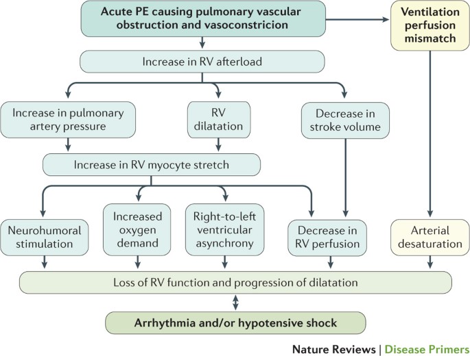
Pulmonary Embolism Nature Reviews Disease Primers
1
1

Pulmonary Embolism Jan Tomis 2018 Pulmonary Embolism Obstruction
1

Pulmonary Embolism In Heart Failure Circulation
Acute Pulmonary Embolism Part 1 Epidemiology And Diagnosis Nature Reviews Cardiology
0 Response to "38 pulmonary embolism pathophysiology diagram"
Post a Comment